The Golgi apparatus or the Golgi body or Golgi complex or simply the Golgi is a complex cytoplasmic structure having relation to cellular secretion. Which consists of vesicles and vacuoles, with smooth cisternae and a network of tubules.
In 1898, Italian neurologist Camillo Golgi observed that Purkinje cells (i.e. nerve cells of the cerebral cortex of the brain) of bran owl contained an internal reticular network that stains black with the silver stain (used silver chromate method). He called this structure apparato reticolare interno (internal reticular apparatus) or Golgi complex.
Various workers have given various names to this complex, such as Dictyosomes, Idiosomes, Lipochondria, Golgi bodies, Golgi substances, Golgi apparatuses, and Golgi complex. Generally, the name Golgi complex is used for the vertebrates and dictyosomes in invertebrates and plants.
Definition of Golgi Apparatus
The Golgi apparatus or the Golgi body or Golgi complex or simply the Golgi is a single membrane-bound flat sac-like and vesicular organelle in the cytoplasm of the eukaryotic cells arranged parallelly in an aggregate and having relation with cellular secretion.
Occurrence
The Golgi apparatus occurs in all cells except the Prokaryotic cells (mycoplasmas, bacteria, and blue-green algae) and Eukaryotic cells of certain fungi, sperm cells of bryophytes, and pteridophytes, cells of mature sieve tubes of plants, and mature sperm and red blood cells (RBC) of animals cells.
In animal cells, there usually occurs a single Golgi apparatus. However, its number may vary from animal to animal and from cell to cell. Thus, the Paramoeba species has two Golgi apparatuses and nerve cells, liver cells, and chordate oocytes have multiple Golgi apparatuses. There are about 50 of them in the liver cells.
Distribution of Golgi Apparatus
In the cells of higher plants, the Golgi bodies or dictyosomes are usually found scattered throughout the cytoplasm and their distribution does not seem to be ordered or localized in any particular manner (Hall et al., 1974).
However, in animal cells, the Golgi complex is a localized organelle. For example, in the cells of ectodermal or endodermal origin, the Golgi complex remains polar and occurs between the nucleus and the periphery (e.g. thyroid cells, exocrine pancreatic cells, and mucus-producing goblet cells of intestinal epithelium) and in the nerve cells, it occupies a circum-nuclear position.
Structure of Golgi Apparatus
The shape of the Golgi apparatus is quite variable (but morphologically similar) in somatic cells of plants and animals. Even in the same cell, there are variations in the functional stage. However, the shape is characteristic of each cell type. In some cell types the Golgi complex appears compact and limited, in others spread out and reticular (net-like).
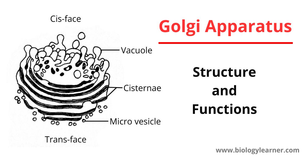
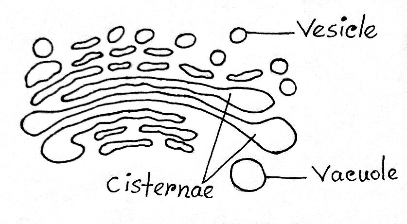
The electron microscopic observations reveal that the Golgi complex comprises Flattened sacs or Cisternae, Large clear vacuoles, Clusters of dense vesicles or Microvesicles, and Tubules.

Flattened Sacs or Cisternae
Cisternae or flattened sacs are flat, elongated, and tubiform membrane-enclosed structures that are closely packed in parallel bundles (stack) lying one above the other. The number of sacs in each Golgi apparatus or complex varies from 3 to 12 in animal cells and it may be up to 30 or more in plant cells.
These sacs (cisternae) consist of double membranes or lamellae, continuous at the two ends, enclosing a thin cavity of about 520 Å. The membranes are mainly lipoproteinous in nature. Each stack of cisternae has a proximal or forming face (cis-face), which is generally convex, and a distal or maturing face (trans-face) of concave shape, which encloses large secretory vesicles.
The cis-face is characterized by the presence of small transition vesicles or tubules that converge upon the Golgi cisternae, forming a kind of fenestrated plate. These transition vesicles are thought to form from the endoplasmic reticulum and then migrate to the Golgi, where they coalesce with each other to form new cisternae.
According to the position of Cisternae, they can be divided into three types:
- Cis-cisternae
- Median cisternae
- Trans-cisternae
In exocrine cells of the pancreas, these sacs are arranged in bundles. Each sac or bundle consists of several membrane pairs separated from other groups by a space (inter-cisternal space) of about 130 Å. Each membrane is 60-70 Å thick and both the membranes of a sac are separated by a space (intra-cistral space) of 60-90 Å wide.
Tubules
Tubules are anastomosing flat networks, arising from the periphery of the cisternae. These flat network tubules are about 300-500 Å in diameter.
Clusters of Dense Vesicles or Microvesicles
Vesicles are small drop-like structures smaller in size about 600 Å in diameter and attached to the tubules or cisternae.
The vesicles are mainly two types:
- Smooth vesicles: These are somewhat flattened and are 200 to 800 Å in diameter and contain secretory material. Thus they are often called secretory vesicles. The smooth vesicles are located within the tubular network or sometimes near the center of the stack of cisternae.
- Coated or rough vesicles: These are spherical outgrowths having about 500 Å in diameter. Their surface is rough and has no known function. The coated or rough vesicles are found at the ends of the tubules or cisternae.
Vacuoles
Vacuoles are large, sac-like globular irregular empty structures situated at the distal ends of cisternae. These arise from trans-cisternae. Vacuoles contain secretory materials of Golgi apparatus. Later, those vacuoles are converted into zymogen granules or lysosomes.
The large vacuoles vary from about 760 Å to about 6000 Å in diameter, whereas in the convoluted tubules, vacuoles range from 500-2000 Å.
The GERL Region
Golgi apparatus is a differentiated portion of the endomembrane system found in both animal and plant cells. This membranous component is spatially and temporally related to the Endoplasmic reticulum (ER) on one side and by way of secretory vesicles, may fuse with specific portions of the plasma membrane.
The trans face of Golgi is associated with the trans-reticular Golgi, TGN (trans-Golgi-network), or GERL (Golgi apparatus + smooth ER + lysosome), in which acid phosphatase enzyme (a characteristic lysosomal enzyme) makes its first appearance.
GERL is found to be involved in the origin of primary lysosomes or zymogen granules and of melanin granules. In the processing, condensing, and packaging of secretory material in endocrine and exocrine cells and in lipid metabolism. GERL is also a region for sorting cellular secretory proteins.
Zones of Exclusion
A Golgi apparatus or Golgi body is surrounded by a differentiated region of cytoplasm where ribosomes, glycogen, and organelles such as mitochondria and chloroplasts are scarce or absent. This region is called the zone of exclusion (Morre et al., 1971) or Golgi ground substance (Sjostrand and Hanzon, 1954).
The endoplasmic reticulum within the zone of exclusion has a smooth surface (lacking ribosomes), and coated vesicles of the Golgi apparatus are restricted to this region. Similar zones of exclusion are associated with microtubules (Porter, 1966), centrioles (Bainton and Farquhar, 1966), and regions of centriole formation (Sorokin, 1968).
Chemical Composition
Chemically, the Golgi apparatus of the rat liver contains about 60% lipid material.
The Golgi apparatus of animal cells contain phospholipids in the form of phosphatidyl-choline. Whereas, that of plant cells contains phosphatidic acid and phosphatidyl-glycerol.
Chemical Properties of Golgi Apparatus
Different parts of the Golgi apparatus have been histochemically identified by specific staining properties, which are:
- Osmium tetroxide (OsO4): This Selectively impregnates the outer face (cis face) of the Golgi apparatus. This stain adheres well to lipids, especially phospholipids and unsaturated fats.
- Phosphotungstic acid (H3PO4. 12WO3. 24H2O): This Selectively stains the maturing or trans face of the Golgi stack. This stain is an anionic stain having a special affinity for polysaccharides and proteins.
- Glycosyl-transferase and thiamine pyrophosphatase: These can be localized cytochemically in the cisternae of the Golgi apparatus. Transferase enzymes are found to be located in the membranes of Golgi, not in the lumen of cisternae (Thorpe, 1984).
- Acid phosphatase: This enzyme is cytochemically marked in the GERL region.
Enzymes in Membranes of Golgi Apparatus
The Golgi apparatus contains a variety of enzymes. Some important enzymes of the Golgi apparatus of animal cells are:
- Glycosyl transgresses:
- Sialyl transferases
- Galactosyl transferases
- Sulpho and glyco-transferases:
- Sulphotransferase
- Lysolecithin acetyltransferase
- Glycero-phosphate phosphatidyl transferase
- Oxidoreductases:
- NADH-cytochrome c-reductase
- NADPH-cytochrome c-reductase
- Phosphatases:
- Glucose-6-phosphatase
- Nucleoside diphosphatase
- ATPase
- Acid phosphatase
- Phospholipases:
- Phospholipase A1
- Phospholipase A2
- Kinases:
- Casein phosphokinases
- Mannosidases:
- Mannosidase I
- Mannosidase II
Functions of Golgi Apparatus
Golgi apparatus appears to play an important role in the storage, packaging, and secretion of certain cell products.
Golgi apparatus performs the following functions of the cell:
- Secretion: The principal function of the Golgi apparatus is secretion. Golgi apparatuses secrete enzymes, hormones, mucus, etc. from secretory cells usually with the involvement of secretory vesicles.
- Absorption of compounds: Golgi apparatus help in absorbing certain compounds like Gold (Au), Copper (Cu), Iron (Fe), etc.
- Lipid transport: Golgi apparatus plays a role in lipid transport. The lipid substance synthesized in the smooth endoplasmic reticulum enters the Golgi complex and is transported to the secretory vesicles.
- Helps in enzyme formation: Golgi apparatus is a great intracellular center of enzyme formation. Golgi complex helps in the production of follicular fluid from granulosa cells of the ovary.
- Production of hormones: Golgi body in endocrine cells helps in the secretion of hormones. Any harm to the Golgi complex in thyroid gland cells will result in a decline in the secretion of its hormone.
- Carbohydrates synthesis: Synthesis of carbohydrate substances from simple sugar may be possible by the involvement of Golgi bodies.
- Storage of protein: Vacuoles and vesicles which are the main components of the Golgi apparatus become filled with protein-lipoid (food materials) material for storage. These stored products help in secretory action.
- Acrosome formation: Acrosome cap of spermatozoan is formed by the direct investment of Golgi apparatuses in the spermatid.
- Formation of lysosome: Golgi apparatus is involved in the formation of the primary lysosome.
- Plant cell wall formation: Golgi bodies during mitotic cell division (at anaphase) form a cell plate at the center of the spindle. This cell plate is gradually enlarged and thickened by the deposition of pectic substances, hemicellulose, and microfibrils of a-cellulose secreted by Golgi bodies.
- Formation of milk protein droplets: In lactating mammary gland of mice are produced protein droplets that are related to the Golgi apparatus. These droplets usually open onto the cell surfaces by fusion of their enclosing membrane with the plasma membrane.
