Mitochondria are the rod-shaped, ovoid, or thread-like, scattered minute particles in all aerobic cells of higher animals and plants. These are also present in algae, protozoa, and fungi. Bacterial cells, however, do not have mitochondria. They are associated with cellular respiration and the source of energy.
In 1850, Kolliker has first observed the mitochondria as granular structures in the striated muscles of insects. Flemming (1882) gave them the name fila. Altmann (1892) first described the mitochondria and he called them bioblasts. While in 1897, Benda coined the term mitochondria. Mitochondria are also known as chondriosomes, plasmosomes, plastochondira, and vermicol.
Definition of Mitochondria
Mitochondria are double membraned spherical, oval, or thread-like auto-replicating cellular organelles present scattered in the cytoplasm of most eukaryotic cells.
Occurrence and Distribution
Mitochondria are absent in prokaryotic cells. However, eukaryotic cells normally contain mitochondria. These are completely lacking in the matured RBC of mammals.
In most cases, mitochondria are uniformly distributed throughout the cytoplasm and move freely in the cytoplasm. Their distribution is somewhat limited in many cells. Mitochondria are present in the basal area of the cell, opposite the renal capillaries, in proximal convoluted tubules of the kidney. They are found between the myofibrils of skeletal muscles.
Structure of Mitochondria
Mitochondria are double membraned, spherical, oval, or thread-like structures uniformly distributed throughout the cytoplasm. Commonly mitochondria are varied in size. They have a length of 1.0-4.1 µm and a diameter of 0.2-1.0 µm (average 0.5 µm).
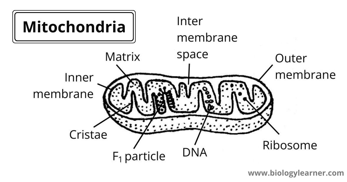
In electron microscopic studies, each mitochondrion consists of mainly four parts- Outer membrane, inner membrane, inner membrane space (peri mitochondrial space), and the matrix.
Outer Membrane
The outside of mitochondria is called the outer membrane.
The outer membrane is made up of protein, lipid, and protein (fluid mosaic in ultrastructure) and is quite smooth. The membrane is composed of approximately 50% of lipid by weight and contains a cusious mixture of enzymes involved in such diverse activities as the oxidation of epinephrine, the degeneration of tryptophan, and elongation of fatty acids.
The outer membrane bears a special type of integral protein called porin that forms large, non-selective membrane chain porin channels through the lipid bilayer. Because of porins, the outer membrane is especially permeable allowing molecules up to about 10000 daltons to freely into the intermembrane space or peri mitochondrial space.
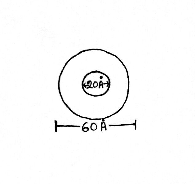
The outer membrane is about 60-70 Å in diameter and bears one type of stalk-less particle called subunits of Parson. The subunit of Parson is a round, hollow structure of 60 Å in diameter. They have a central hole of 20 Å in diameter.
Intermembrane Space or Peri Mitochondrial Space
The space between the outer and inner membranes of mitochondria is called intermembrane space or peri mitochondrial space.
This space is filled with watery fluid and is 40-70 Å in width. This space may be designated into two regions- The peripheral space (located near the periphery), and intra cristal space.
Very few enzymes are present in this space, like- NADH dehydrogenase, succinate, cytochrome oxidize, etc.
Inner Membrane
The inner side of mitochondria is called the inner membrane.
This membrane also consists of protein, lipid, and protein (fluid mosaic in ultrastructure) but lacks porin protein as in the outer membrane. The inner membrane has an outer cystol face or C-face towards the inter mitochondrial space and an inner matrix face or M-face.
The inner membrane is thrown up into a series of folds called Cristae (animals) or tubuli or microvilli (plants) which project into the inner membrane. The cavity of the cristae is called inter cristal space.
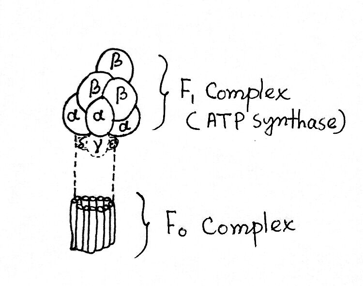
A special type of lipid cardiolipin is present only in the inner membrane. In the M-face, the inner membrane bears many tennis racket-like staked round bodies which are called oxysomes or subunits of Farnandez-Moran or elementary particles or F1 particles.
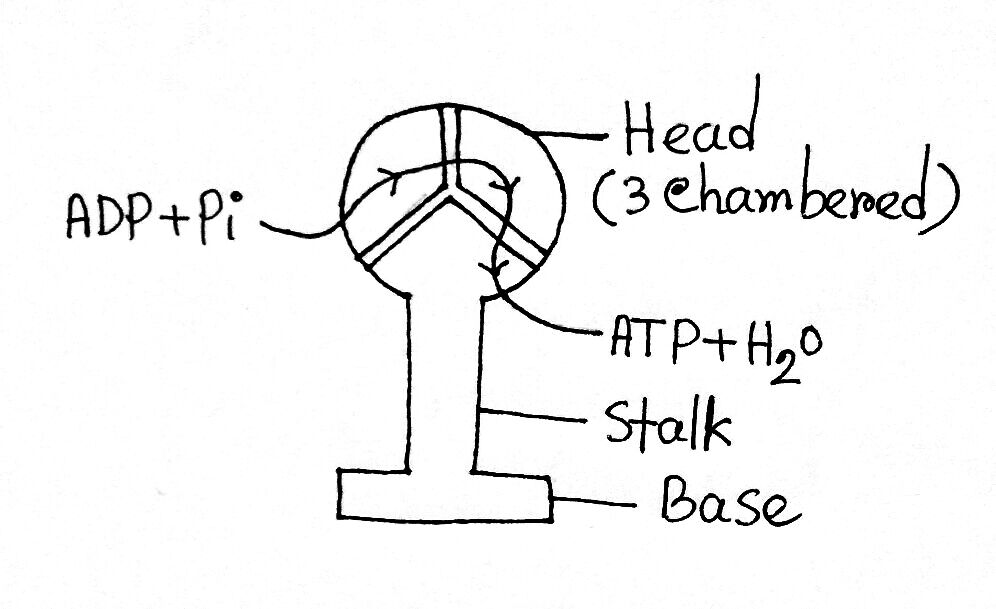
The oxysome is made up of three parts:
- One round head, which has 3 segments (75-100 Å in diameter)
- A long stalk (50 Å in length)
- A rectangular base is inserted into the membrane (110 Å in length and 15 Å in diameter).
The ADP and inorganic Pi enter through the 3 segments of the head (f0-f1 complex) and give rise to the ATP and H2O molecules.
Mitochondrial Matrix
Internal space enclosed within the inner membrane of mitochondria is called the mitochondrial matrix. The matrix is the site of the Krebs cycle and bears many enzymes like- dehydrogenase and the essential enzymes of DNA synthesis, RNA synthesis, and protein synthesis.
The matrix contains its own circular double helix DNA (mt-DNA), RNA, and ribosomes.
Mitochondrial DNA
Mitochondrial DNA is double helix and circular in nature and is different from that nuclear DNA. It is made up of one heavy strand and one light strand. Mitochondrial DNA does not contain histone proteins and has no nucleosome. It occurs in multiple copies and most usually remains attached to the inner mitochondrial membrane. Repetitive sequences are rare in this type of DNA. Mitochondrial DNA is inherited maternally.
Mitochondrial Ribosome
The ribosome of mitochondria is called the mitoribosome. This type of ribosome is smaller in diameter than the cytoplasmic ribosome. Mitochondrial ribosomes require a higher concentration of Mg2+ ions to maintain their integrity than do cytoplasmic ribosomes.
It is 78S, 75S, 73S, and 60S types.
- 60S (Human)→ (45+35)S
- 75S (Yeast)→ (53+35)S
- 73S (Neurospora sp.)→ (50+37)S
Mitochondrial RNA
Mitochondrial RNA (mRNA), which is distinct from RNA of nuclear origin, has been found in its mitochondrial fraction.
Functions of Mitochondria
Mitochondria are the powerhouses of the cell. They supply nearly all the required biological energy. The mitochondria are fully capable of converting pyruvic acid to CO2 and H2O.
The functions of mitochondria are as follows:
- Cell respiration: Krebs cycle is the one main phase of cell respiration sited inside of the mitochondrial matrix and produces 30 molecules of ATP from 2 molecules of pyruvic acid (Acetyl-CoA→Oxaloacetic acid→Citric acid).
- ATP transport: The ATP molecules produced as of cellular respiration accumulate in the mitochondria. The mitochondria are collected at sites where energy is hydrophobic. As a result of membrane concentration and an increase in internal hydrostatic pressure of the mitochondria, water and ATP are squeezed out. This results in a lowering of the ATP concentration and mitochondrial membrane relaxation. The thyroid hormone thyroxine causes the mitochondria to swell, while ATP brings about concentration.
- Lipid synthesis: A set of enzymes that control the synthesis of lecithin and phosphatidyl-ethanolamine from fatty acids, glycerol, and nitrogen bases are present in most of the mitochondria.
- Elongation of fatty acids: Mammalian mitochondria have a group of enzymes that carry out the elongation of fatty acids by adding acetyl-CoA and subsequently reducing the keto acid produced. By this method, myristate is elongated to palmitate(palmitic acid), palmitate to stearate(Stearic acid), etc.
