Ribosomes are membraneless, small, sub-spherical ribonucleoprotein particles.
- Ribosomes are often found connected to the outside of the endoplasmic reticulum and nucleus.
- They also occur freely in the cytoplasm (cytoplasmic matrix), chloroplasts, and mitochondria.
- In 1943, A. Claude has first isolated the ribonucleoprotein particles (the ribosomes) from the cytoplasm. He named these ribonucleoprotein particles as microsomes (Claude’s particles).
- During the microscopic study of bean roots, the ribosomes were first observed in plant cells by Robinson and Brown in 1953. Later in 1955, G. E. Palade observed them in animal cells and termed them ribosomes (also called Palade’s particles).
Definition of Ribosomes
Ribosomes are small, dense, membraneless, rounded, and granular ribonucleoprotein organelles which occur either freely in the matrix of mitochondria, chloroplast, and cytoplasm or remain attached to the membranes of the endoplasmic reticulum and nucleus.
Occurrence
The ribosomes occur in both Prokaryotic cells and Eukaryotic cells. In prokaryotic cells, the ribosome often occurs freely scattered in the cytoplasm. In eukaryotic cells, the ribosome either occurs freely in the cytoplasm (e.g.- yeast cells, lymphocytes, meristematic plant tissues, embryonic nerve cells, etc.) or remains attached to the outer surface of the membrane of the endoplasmic reticulum and nucleus (e.g.- pancreatic cells, plasma cells, hepatic parenchymal cells, serous cells, Nissls bodies, etc). The cells in which active protein synthesis occurs, ribosomes attach to the membranes of the endoplasmic reticulum.
When the ribosomes are not attached to the endoplasmic reticulum, they are called free ribosomes. Free ribosomes serve as sites for the synthesis of proteins that are required for intracellular utilization and storage.
Number of Ribosomes
Ribosomes are innumerable in a cell, especially in endoplasmic reticulum-containing cells. A single cell of E. coli contains 20000-30000 ribosomes.
In yeast cells, at the base of gland cells, in plasma and liver cells, in nerve cells, and in all rapidly growing plant and animal cells, they are in large numbers.
Types of Ribosomes
The size, volume, structure, composition, etc. of the ribosome of an organism remains the same in different types of cells. Based on the size and Sedimentation coefficient (S), Ribosomes are basically two types: 70S ribosomes and 80S ribosomes.
The sedimentation coefficient is expressed in the Svedberg unit i.e., the S unit. Svedberg units are not directly additive, they represent a rate of sedimentation, not weight. S= 1×10-13 cm/s/dyne/gm
70S Ribosome
They are relatively smaller in size and have a sedimentation coefficient of the 70S. This type of ribosome consists of a large 50S subunit and a small 30S subunit.
They occur in the prokaryotic cells (e.g., bacteria, blue-green algae) and also in mitochondria and chloroplasts of eukaryotic cells.
The molecular weight of the 70S ribosome is 2.5×10⁶ daltons and the dimension of each ribosome is about 200-290 Å × 170-210 Å.

80S Ribosome
They are relatively larger in size than 70S ribosomes and are mainly found in the cytoplasm of eukaryotic cells (plant and animal cells).
The sedimentation coefficient of this type of ribosome is the 80S. Each 80S ribosome is made up of two subunits the smaller 40S subunit remains attached to a larger 60S subunit like a cap. Their molecular weight is 2.7 × 10⁶ daltons and the dimension of 300-340 Å × 200-240 Å.
Other Forms of Ribosomes
The ribosomes are also divided into the following types based on their location:
- Bacterial ribosome: This type of ribosome is found in bacterial cells and it has a sedimentation coefficient of the 70S.
- Mitochondrial ribosome: The ribosomes which are found in mitochondria are called mitoribosomes. They are many types, like- the mitoribosomes of fungus are 73S type, plants have 78S type of mitoribosomes (e.g.- Corn), while the mitoribosomes of mammals are of the 60S. However, mitoribosomes are generally considered to be of the 70S.
- Plastidial ribosome: The ribosomes found in chloroplasts are called plastidial ribosomes or plastidoribosomes. The plastidoribosomes are the 70S in the higher plants.
- Nuclear ribosome: The ribonucleoprotein particles found in the nucleus are called nuclear ribosomes.
- Nucleolar ribosome: The RNP (ribonucleoprotein) particles of 150-200 Å in diameter found in the nucleolus are called nucleolar ribosomes.
- Cytoplasmic ribosome: The large ribosome particles of the cytoplasm are called cytoplasmic ribosomes.
Structure of Ribosomes
Ribosomes are oblate spheroid structures and the most abundant organelles of a cell. Each ribosome is about 250 Å in diameter.
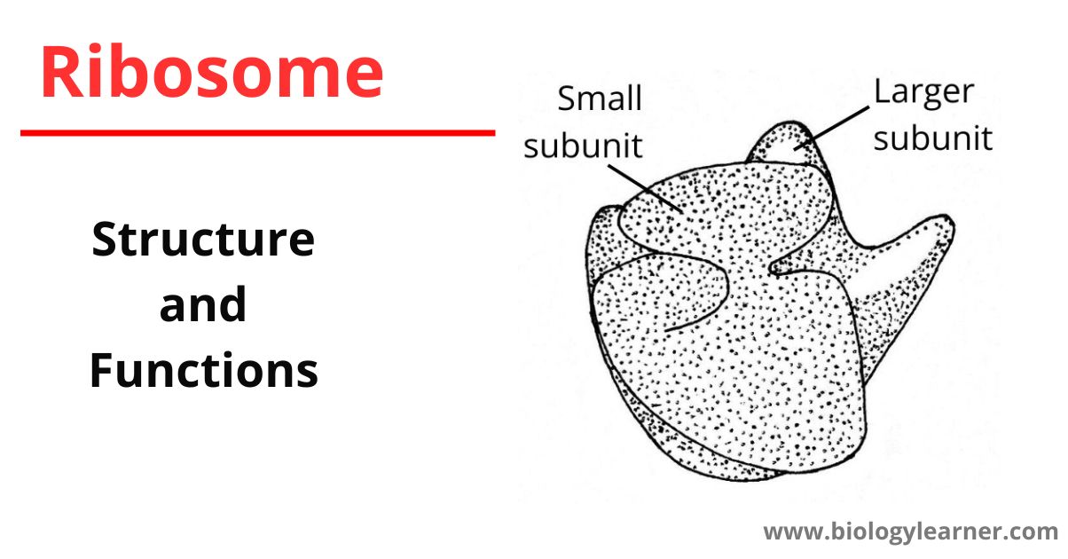
In the electron microscopic studies, negative staining reveals that each ribosome is porous, hydrated, and composed of two unequal sub-units, one is larger and the other is a smaller subunit. The larger subunit is dome-shaped or cup-shaped (140-160 Å), while the smaller one is oblate-ellipsoid (90-110 Å). The smaller subunit is about half the size of the larger subunit and occurs above the larger subunit to form a cap-like structure.
Each 70S ribosome consists of two subunits as 50S and 30S. The larger 50S subunit has a molecular weight of 1.8 × 10⁶ daltons, while the molecular weight of the smaller 30S subunit is about 0.9 × 10⁶ daltons. On the other hand, the 80S ribosome also consists of two subunits as 60S and 30S. The molecular weight of the 60S subunit is 1.5-1.8 × 10⁶ daltons and 3-3.5 × 10⁶ daltons in the 40S subunit.
The two structural (cup and cap) ribosomal subunits remain united with each other due to the high concentration (0.001M) of the Mg2+ ions. When the concentration of Mg2+ ions reduces in the matrix of the cytoplasm, both subunits get separated. At a high concentration of Mg2+ ions in the cytoplasmic matrix, the two ribosomes become associated with each other to form the dimer.
Polyribosome: During protein synthesis, many ribosomes bind to an individual mRNA (messenger RNA) molecule forming a polyribosome or polysome or ergosome.
The ribosomes are composed of highly flooded ribosomal RNA(rRNA) and many attached proteins. The RNA and proteins are intertwined and arranged in a complex manner in the form of two subunits.
Various models have been introduced regarding the structure of ribosomes. Among them, J. A. Lake given asymmetrical model (also known as Lake’s model) is the most common and universally accepted.

According to this model, the smaller subunit has three parts- a head, a base, and a platform. The platform separates the head from the base by a cleft. On the other, the larger subunit also consists of three parts- a ridge, a central protuberance, and a stalk. The first two are separated with the help of a valley.
Three special regions are seen between the two subunits of a ribosome. These are aminoacyl site (A-site) or acceptor site, peptidyl site (P-site), and exit side (E-site). During the protein synthesis, aminoacyl-tRNA bounded to the ribosome at the A-site, peptidyl-tRNA bounded to the ribosome at the p- site, and while at E-site, the deacylated-tRNA exits from the ribosome.
Chemical Composition of Ribosomes
The major constituents of ribosomes are highly flooded ribosomal RNA (rRNA) and proteins. The lipids are entirely absent or present in traces.
Ribosomal RNAs
More than half of the weight of the ribosome is ribosomal RNA(rRNA). In prokaryotes, the ribosomes contain 66% of rRNA and eukaryotic ribosomes contain 60% of rRNA.
The 70S ribosomes contain three types of rRNA:
- 23S rRNA (3300 nucleotides)
- 16S rRNA (1650 nucleotides)
- 5S rRNA (120 nucleotides)
The larger 50S ribosomal subunit contains 23S and 5S rRNA, while the 16S rRNA occurs in the smaller 30S subunit.
The 80S ribosomes possess four types of rRNA:
- 28S rRNA (4700 nucleotides)
- 18S rRNA (1900 nucleotides)
- 5.8S rRNA (160 nucleotides)
- 5S rRNA (120 nucleotides)
The 28S, 5S, and 5.8S rRNAs are present in the larger 60S ribosomal subunit and the smaller 40S subunit contains 18S rRNA.
Ribosomal Proteins
The ribosomal proteins may be basic, structural, or enzymatic in function. The ribosomal proteins of smaller ribosomal subunits are termed S-proteins, while L-proteins are in larger ribosomal subunits.
The larger ribosomal subunit contains an important enzyme – peptidyl transferase which catalyzes the formation of the peptide bonds. Inside the ribosome, the rRNA remains fully adhered to proteins. The ribosomes are, therefore, ribonucleoprotein(RNP) particles.
Metallic Ions
In addition to rRNA and proteins, ribosomes also contain some divalent metallic ions, such as Mg2+, Ca2+, and Mn2+.
Functions of Ribosomes
- Protein synthesis: The principal function of a ribosome is protein synthesis(translation-process of synthesizing proteins). In all living cells, ribosome serves as the site of biological protein synthesis.
- Transport the synthesized proteins: Ribosomes also function as the transporter of the synthesized proteins.
- Helps in protoplasm formation: The free ribosomes present in the cytoplasm synthesize various proteins that help in the formation of protoplasm.
- As catalysts: Ribosome acts as catalysts in the biological processes of peptidyl transfer and peptidyl hydrolysis.
- Plays a protective role: The newly synthesized polypeptide chains passing through the tunnel or channel of the larger subunit of the ribosome are protected against the action of protein-digesting enzymes.
References
- Wilson, D. N., & Doudna Cate, J. H. (2012, May 1). The Structure and Function of the Eukaryotic Ribosome. Cold Spring Harbor Perspectives in Biology, 4(5). https://doi.org/10.1101/cshperspect.a011536
- Verma, P. S., & Agrawal, V. K. (2006). Cell Biology, Genetics, Molecular Biology, Evolution & Ecology (1 ed.). S .Chand and company Ltd.
- Spirin, A. S. (2002, January 25). Ribosome as a molecular machine. FEBS Letters, 514(1), 2–10. https://doi.org/10.1016/s0014-5793(02)02309-8
- Opron, K., & Burton, Z. (2018, December 21). Ribosome Structure, Function, and Early Evolution. International Journal of Molecular Sciences, 20(1), 40. https://doi.org/10.3390/ijms20010040
- Ribosome Structure. (2019, April 3). News-Medical.net. https://www.news-medical.net/life-sciences/Ribosome-Structure.aspx

Helpful info.
This is really interesting, You’re a truly highly trained author. Nice post…
Excellent post.
Your article is truly informative.
Great article and straight to the point.
The information you have was perfect that we’re seeking and i also really enjoyed the content. I would like to visit your site again from now on.
Nice to see it helps you.