The endoplasmic reticulum (ER) is a tubular branched cell organelle in the cytoplasm of the Eukaryotic cell that divides the cytoplasm into several compartments. They are extended from the nuclear membrane to the plasma membrane.
Porter (1945) has observed the endoplasmic reticulum in electron micrographs of liver cells. But in 1953, he coined the name endoplasmic reticulum for this organelle.
All eukaryotic cells contain endoplasmic reticulum in the endoplasm or cytoplasm. The erythrocytes (RBC), egg, and embryonic cells are lacking in the endoplasmic reticulum.
Definition of Endoplasmic Reticulum
The endoplasmic reticulum is the tubular organelle that remain within the cytoplasm to divide the cytoplasm into irregular compartments.

Origin
At present, the manner of origin of the endoplasmic reticulum is not definitely known. The most explicit hypothesis is that the endoplasmic reticulum is “budded” off from the nuclear membrane (Wischnitzer, 1974).
The ER appears to arise from the outer membrane of the nuclear membrane by out folding, or from the plasma membrane by in folding. The smooth ER seems to arise from the rough ER by the detachment of ribosomes.
Structure of Endoplasmic Reticulum
The endoplasmic reticulum is bounded by a thin single membrane of 50-60 μm thickness. The cavity of the ER is well developed and acts as a passage for secretory products. These cavities are full of fluid called the endoplasmic matrix.
Structurally, the ER may occur in the following forms: Cistarnae (lamellar form), Vesicle (vesicular form), and, Tubules (tubular form).
Cisternae
The cisternae are long, flattened, sac-like, unbranched tubules having a diameter of 40-50 μm. They are arranged parallelly in bundles or stakes within the cytoplasm.
Sometimes ribosomes are associated with their membrane.
Vesicles
The vesicles are oval, membrane-bound vascular structures having a diameter of 25-500 μm. They often remain isolated in the cytoplasm and occur in most cells.
Vesicles are also called Microsomes.
Tubules
The tubules are branched structures forming the reticular system along with the cisternae and vesicles. The dimension of the tubules varies from 50-190 μm and occurs almost in all the cells.
The membrane of the tubules may also be associated with ribosomes.
Types of Endoplasmic Reticulum
Based on the presence or absence of ribosomes on the surface of the ER, they may be classified into two types- Agranular or Smooth Endoplasmic Reticulum (SER) and Granular or Rough Endoplasmic Reticulum (RER).
Smooth Endoplasmic Reticulum
This type of ER possesses smooth walls because the ribosomes are not attached to its membranes. So, the ER without ribosomes on its surface is called Smooth Endoplasmic Reticulum.
The smooth type of ER occurs mostly in those cells, which are involved in the metabolism of lipids (including steroids) and glycogen. SER is generally found in adipose cells, interstitial cells, glycogen storing cells of the liver, conduction fibers of the heart, spermatocytes, and leukocytes. The presence of the smooth type of ER in the muscle cells is called the Sarcoplasmic Reticulum. In the pigmented retinal cells it exists in the form of tightly packed vesicles and tubes known as myeloid bodies.

Although the SER forms a continuous system with RER, it has different morphology. For example, liver cells consist of a tubular network that pervades a major portion of the cytoplasmic matrix. These tubules are present in regions rich in glycogen and can be observed in the matrix as dense particles, called glycosomes.
Glycosomes measure 50-200 nm in diameter and contain glycogen along with enzymes involved in the synthesis of glycogen (Rybicka, 1981). Many glycosomes attached to the membranes of SER have been observed by electron microscopy in the liver and conduction fiber of the heart.
Rough Endoplasmic Reticulum
The granular or rough type of ER possesses rough walls because the ribosomes remain attached to its membranes. So, the membrane of the ER contains ribosomes attached to its surface, called Rough Endoplasmic Reticulum (RER).
The rough ER is found abundantly in those cells which are active in protein syntheses such as pancreatic cells, plasma cells, goblet cells, and liver cells.

The rough type of ER takes basophilic stain due to its RNA contents of ribosomes. The region of the matrix containing the rough type of ER takes basophilic stain and is named ergastoplasm, basophilic bodies, chromophilic substance, or Nissl bodies by early cytologists.
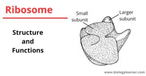
In the rough ER, ribosomes are often present as polysomes held together by mRNA and are arranged in typical rosettes or spirals. RER contains two transmembrane glycoproteins, called Ribophorin (ribosomes containing protein). Ribophorin I is the glycoprotein of about 65000-dalton molecular weight. While Ribophorin II is the glycoprotein of 64000-dalton molecular weight. These ribophorins are attached to the ribosomes by their 60S subunits.
Enzymes of the ER Membranes
The membranes of the ER are found to contain many kinds of enzymes that are needed for various important synthetic activities.
Some of the most common enzymes are:
- Stearases
- NADH-cytochrome C reductase
- NADH diaphorase
- Mg2+ activated ATPase
- Glucose-6-phosphatase
- 5′-nucleotidase
- Acyle transferase
- Colin phosphotransferase
Certain enzymes of the ER such as nucleotide diphosphate are involved in the biosynthesis of the phospholipid, ascorbic acid, steroids, glucuronide, and hexose metabolism.
Functions of the enzymes of ER Membranes
- Synthesis of glycerides (e.g. triglycerides, phospholipids, glycolipids, and plasmalogens)
- Metabolism of plasmalogens
- Synthesis of fatty acids
- Biosynthesis of the steroids (e.g. cholesterol biosynthesis, steroid hydrogenation of unsaturated bonds
- NADPH2 + O2-requiring steroid transformations: Aromatization and hydroxylation.
- NADPH2 + O2-requiring steroid transformations: Aromatic hydroxylations, side-chain oxidation, deamination, thioether oxidation, desulphuration
- L-ascorbic acid synthesis
- UDP-uronic acid metabolism
- UDP-glucose dephosphorylation
Functions of Endoplasmic Reticulum
The endoplasmic reticulum acts as the secretory, storage, circulatory and nervous system for the cell.
It performs the following important functions:
Common Functions of Endoplasmic Reticulum (SER and RER)
- Mechanical support: The ER provides an ultrastructural skeletal framework to the cell and gives mechanical support to the colloidal cytoplasmic matrix.
- Exchange of ions and any other fluid: The exchange of molecules by the process of osmosis, diffusion, and active transport occurs through the membranes of ER. Like the plasma membrane, the ER membrane has permeases and carriers.
- Perform various metabolic activities: The endoplasmic membranes contain many enzymes which perform various synthetic and metabolic activities. Further, the ER provides an increased surface for various enzymatic reactions.
- Isolation of metabolic products: ER keeps the metabolic byproducts in isolation and separates the organic reactions in the cell cytoplasm. In this way, ER controls the osmotic pressure in a cell.
- Intra-cellular transport: The ER acts as an intra-cellular circulatory or transporting system. Various secretory products of granular ER are transported to various organelles as Granular ER→agranular ER→Golgi membrane→lysosomes, transport vesicles, or secretory granules. Membrane flow may also be an important mechanism for carrying particles, molecules, and ions into and out of the cell. Export of RNA and nucleoproteins from the nucleus to the cytoplasm may also occur by this type of flow.
- Intra-cellular impulse conduction: The ER membranes are found to conduct intra-cellular impulses. For example, the sarcoplasmic reticulum transmits impulses from the surface membrane into the deep region of the muscle fibers.
- Inter-cellular connection: Inter-cellular connection of ER between two adjacent cells is maintained by desmotubules.
- Formation of nuclear envelope: The ER membranes form the new nuclear envelope after each nuclear division.
- Formation of cell organelles: Most of the cell organelles like Golgi bodies, mitochondria, lysosomes, cell plates, etc. are usually developed from the ER.
- Release of calcium: The sarcoplasmic reticulum plays a role in releasing calcium when the muscle is stimulated and actively transporting calcium back into the sarcoplasmic reticulum (When the stimulation stops and the muscle must be relaxed).


