Firstly cyanobacteria are kept in as myxophyceae of class algae (blue-green algae). Later some phycologists kept it into Cyanophyceae, while others kept it into Schizophyceae also. On the basis of the electron microscope and biochemistry Echlin and Moris that cyanobacteria show great affinity with blue-green algae.
Under the rules of the International Code of Nomenclature of Bacteria (1978), blue-green algae have been proposed to be named Cyanobacteria by some bacteriologists.
According to Kunisawa, Mandel, Cohn, and Bezire, the organisms of cyanobacteria are not algae and they form a major group of bacteria. The group of blue-green algae, which are chlorophyllous and photosynthetic is called cyanobacteria.
Greek words “cyano” = blue and “bact” = rod
The absence of a definite nucleus with typical chromosomes, and the distribution of the pigments in a primitive chromatophore structure, show their close resemblance with bacteria.
Definition of Cyanobacteria
Cyanobacteria are the oxygenic photosynthetic bacteria growing on the surface of freshly exposed rocks, resulting in the deposition of organic matter due to the accumulation of their cells.
Salient Features of Cyanobacteria
Their major characteristics features are:
- Its members are blue in color due to the presence of phycocyanin pigment, like; chlorophyll-a, carotene, and phycoerythrin also occur.
- Pigments are distributed in the cytoplasm instead of chromatophores.
- The true nucleus is absent.
- The nuclear membrane and nucleolus are absent.
- Cell divisions occur through protoplasm division and septum formation.
- Reserve food glycogen is similar to cyanophycean starch.
- Reproductive units are non-flagellate, hence non-motile.
- Sexual reproduction is completely absent.
- The mucilaginous or gelatinous sheath is present on the thallus.
- Pyrenoids are absent.
- These are prokaryotes, which lakes plastids, vacuoles, mitochondria, Golgi body, and endoplasmic reticulum.
- The cell wall is without cellulose.
- The cell wall contains 8 amino acids with peptides and diaminopimelic acid and glucosamine.
Similarities between Cyanobacteria(blue-green algae) and Bacteria
- Presence of a prokaryotic nucleus.
- Absence of well-organized plastids.
- Presence of sheaths.
- Ability to fix atmospheric nitrogen.
Differences between Cyanobacteria and Bacteria
- Chlorophyll-a is absent in bacteria.
- Magnesium porphyrin compound is present in photosynthetic bacteria which is similar to chlorophyll-a.
- Heterocyst is absent in bacteria.
- Photosynthetic bacteria produced Oxygen.
Examples
The few common genera of blue-green algae are Aulosira, Nostoc, Stigonema, Anabaena, Spirulina, Oscillatoria, Syctonema, Gloeocapsa, Chrococcus, etc.
Thallus Organization in Cyanobacteria
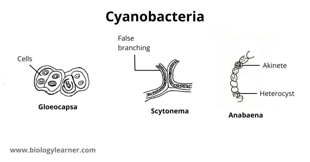
In members of cyanobacteria, the thallus structure is very simple. Some members like Chroococcus, Gloeocapsa, and Chamaesiphon are unicellular. Other forms like Microcystis and Merismopedia are colonial and their colony may be irregular, square-like, or spherical.
The thallus of some members may be filamentous as in Oscillatoria, Lyngbya, Anabaena, Nostoc, Scytonema, Rivularia, and Gloeotrichia. Their filaments remain surrounded by a mucilaginous sheath.
When cells are arranged in a linear row, the structure is called a trichome. In the case of Oscillatoria, the breadth of the trichome is uniform but in Rivularia, the anterior part of the trichome is pointed.
The trichome of Nostoc is unbranched, while in Nostochopsis, the trichome is branched. There occurs false branching in the filament of Scytonema.
Cell Structure of Cyanobacteria
With the help of an electron microscope, Pankratz and Bowen (1963) studied the structure of cyanobacterial cells. The detailed cell structure of cyanobacteria is as follows:
Cell Sheath
The cyanobacterial cell is surrounded by a hygroscopic gelatinous sheath, which is made up of three layers of microfibrils. The microfibrils are composed of pectic acids and mucopolysaccharides. Sheaths are reticulately arranged within a matrix to give a homogeneous appearance.
The function of the cell sheath is to absorb and retain water.
Cell Wall
A multi-layered wall is present below the gelatinous sheath. The cell wall is made up of four layers which are designated numerically as LI, LII, LIII, and LIV.
The layers LI and LIII are electron transparent, while LII and LIV are electron-dense.
- Layer LI: The innermost layer of the cell wall is LI. It is present next to the plasma membrane. The layer LI is of about 3-10 nm thickness and is enclosed by the layer LII.
- Layer LII: LII is an electron-dense, thin layer. It is about 10-1000 nm in thickness. The layer LII consists of muramic acid, glutamic acid, alanine, glucosamine, and α-di-amino-pamelic acid.
- Layer LIII: The electron transparent Layer III is of about 3-10 nm thickness, just like layer I.
- Layer LIV: The outermost layer is LIV which is a thin and again electron-dense layer.
Plasma Membrane
Below the cell wall, a thin cytoplasmic bilayer membrane called the plasma membrane is present. The plasma membrane consists of two electron-opaque layers separated by a translucent layer.
It has a thickness of 7 nm, is selectively permeable, and maintains the physiological integrity of the cell.

Cytoplasm
The cytoplasm is differentiated into two regions, the outer colored peripheral region which is called the chromoplasm, and the central colourless region called the centroplasm or central body.
Chromoplasm
The chromoplasm contains flattened vesicular structures called photosynthetic lamellae or thylakoids.
- Lamellae: In the peripheral part of the cyanobacterial cell (in chromoplasm), many lamellae or thylakoids are present. The lamellae are arranged in two or more parallel stacks or scattered irregularly. Each lamella is represented by a flat sac, which is surrounded by a unit membrane. Each unit membrane is about 75 Å in thickness. These lamellae are separated by a space of 50 μ. This space is occupied by many rows of discoidal phycobilisomes, which contain phycocyanin pigment and, to a lesser extent phycoerythrin pigment.
Centroplasm or Central Body
The central body, or centroplasm, is colorless and irregular in shape, occupying more or less a quarter or one-third of the total volume of the cell.
Some people are of the opinion that the centroplasm is the storehouse of food and according to others, it is an incipient nucleus or nucleoplasm.
The nucleoplasm contains numerous fine, randomly oriented DNA fibrils.
Cytoplasmic Inclusions
In a typical cyanobacterial cell, many sub-cellular organelles such as cyanophycean granules, gas vacuoles, polyglucoside bodies, ribosomes, polyhedral bodies, polyphosphate bodies, etc. are present.
- In many cyanobacteria (e.g., Anabaena, Gloeotrichia, Microcystis, Oscillatoria, etc.), pseudovacuoles (pockets of gas) are found to be distributed throughout the cytoplasm. These pseudovacules provide greater buoyancy in such species and also act as light screens against intense illumination.
- The proteins present in the cell are synthesized by ribosomes.
- Cyanophycean granules or cyanophycin store reserve food material.
- Polyphosphate bodies are meant for the storage of phosphorus.
- Polyhedral bodies are also called carboxysomes. They produce an important enzyme called ribulose diphosphate carboxylase.
- Other granules are called storehouses of nitrogen.
Economic Importance of Cyanobacteria
- Cyanobacteria are one of the early colonizers of bare and barren areas. They provide suitable conditions for the growth of other organisms.
- Many blue-green algae have the ability of nitrogen fixation. The filamentous forms possess heterocysts for fixing nitrogen. Some of the fixed nitrogen comes out as excretion. After the death of the algae, the substratum becomes rich in nitrogen.
- Nitrogen fixing Cyanobacteria are often used for the reclamation of usar soils, e.g., Anabaena, Nostoc. They produce acidic chemicals for counteracting the alkalinity of the soil and nitrogenous compounds that are generally deficient in these soils.
- Cyanobacteria function as food for many aquatic animals. Spirulina is used as a nutritional supplement, rich in protein, vitamins, and minerals for humans and animals. Nostoc and Anabaena are also used as fodder and manure.
- Lyngbya serves as a source for the production of antibiotics.
- Cyanobacteria produce water blooms, imparting bad odour and colour to water bodies.
- They can grow on the walls and roofs of buildings during the rainy seasons, causing discoloration, corrosion, and leakage.
- Some Cyanobacteria (e.g., Microcystis aeruginosa, Anabaena flos-aquae, Aphanizomenon flos-aquae) produce toxins that are harmful to most aquatic animals. They may prove equally toxic to human beings drinking or bathing in such water.

Nice😊