Marchantia, a genus of thallose liverworts in the order Marchantiales, is widely distributed all over the world.
- It belongs to the family Marchantiaceae.
- Nicolas Marchant, a French botanist, named the Marchantia in memory of his father.
| Division: | Bryophyta |
| Class: | Hepaticopsida |
| Order: | Marchantiales |
| Family: | Marchantiaceae |
| Genus: | Marchantia |
Distribution of Marchantia
Marchantia is the most prominent genus in the family Marchantiaceae. The genus is represented by about 65 species and is found in all parts of the world.
All the species are terrestrial and prefer to grow in moist and shady places like wet open woodlands, banks of streams, wood rocks or on shaded stub rocks. A few species occur (e.g., Marchantia polymorpha) as pioneers in burnt forest soil.
- Some of the common Marchantia species are Marchantia indica, M. polymorpha, M. palmata, M. nepalensis, etc.
Gametophyte of Marchantia
Like that of Riccia, the gametophyte of Marchantia is a thallus. The haploid gametophytic thallus is the dominant phase of the life cycle.
Morphology of Marchantia
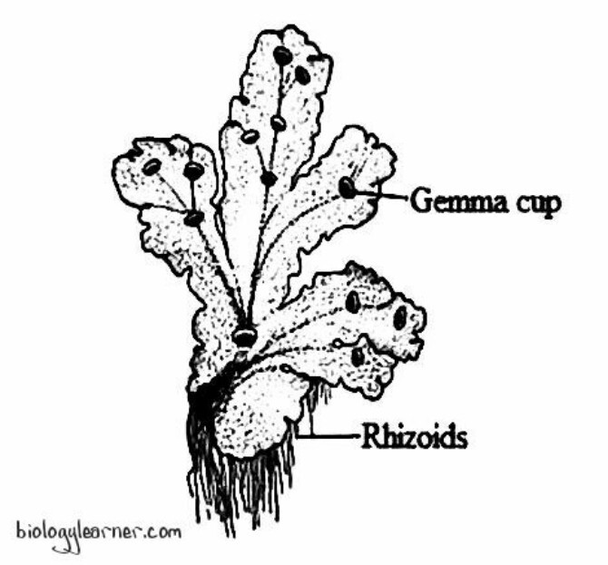
The plant body of Marchantia is gametophytic. The gametophyte is a flat, prostrate, dorsiventral, dichotomously branched thallus.
Dorsal Surface
The dorsal surface of the Marchantia thallus is dark green, with each branch having a median, more or less conspicuous midrib. The midrib in each branch of the thallus is marked on the dorsal surface by a shallow groove and on the ventral surface by a low ridge.
The dorsal surface of the thallus has a number of regular polygonal or rhomboidal areas called areolae. These polygonal areas demarcate the outline of the underlying air chamber. Each polygonal area has a pore at the centre called an air pore.
Each branch of the thallus has a growing point situated at the apex of a terminal groove called an apical notch.
The dorsal surface of the mature thallus bears vegetative and sexual reproductive structures. The vegetative reproductive structures are gamma cups, which develop along with the midrib. The gamma cups are cup-like or crescent-shaped structures with spiny or fimbriate margins.
Sexual reproductive structures (sex organs) are borne on special erect branches called gametophores or gamctangiophores. These branches are umbrella-shaped and arise from the distal end of the thallus at the growing point (apical notch).
The gametophores bearing antheridia (male sex organs) are called antheridiophores, and those bearing archegonia (female sex organs) are called archegoniophores. Marchantia is a heterothallic (dioecious) plant. Therefore, a thallus bears either antheridiophores or archegoniophores.
Ventral Surface
The ventral surface of the Marchantia thallus bears numerous scales and rhizoids along the midrib.
Scales
The scales are multicellular, one-cell thick, and usually violet-coloured structures that increase the surface area for absorption. The colour of the scale is due to the presence of anthocyanin pigments. These scales are arranged in 3–4 rows on both sides of the midrib.
Scales mainly protect the growing point and retain water by capillary action.
Scales are of two types: appendiculate and ligulate.
- Appendiculate scales: The appendiculate scales are larger and more elaborate due to the presence of an apical sub-rounded appendage. They form the inner row of the scales, close to the midrib.
- Ligulate or simple scales: The ligulate scales are relatively smaller than the appendiculate scales and without appendages. They form the outer or marginal row.
Rhizoids
The rhizoids are unicellular, branched, colourless structures present between the scales on the ventral surface of the thallus. They develop as prolongations of the lower epidermal cells.
The main functions of rhizoids are to attach the thallus to the substratum and to absorb water and mineral nutrients from the soil.
Rhizoids are of two types: smooth-walled rhizoids and tuberculate rhizoids.
- Smooth-walled rhizoids: The smooth-walled rhizoids are simple, elongated, and slightly wider. They contain a smooth inner wall with colourless contents.
- Tuberculate or pegged rhizoids: Tuberculate rhizoids are thinner and have peg-like or plate-like projections in the cell lumen. They appear like circular dots on the surface view.
Anatomy of Marchantia
Internal morphology, or internal structure of the thallus, is the anatomy of the Marchantia thallus.
In a vertical cross-section, the Marchantia thallus shows an internal differentiation of tissues distinctly organised into three regions: the epidermal region, the photosynthetic region, and the storage region.
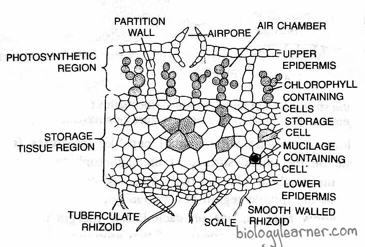
Epidermal Region
The epidermal region is composed of well-defined upper and lower epidermis. The epidermis is formed of thin-walled, quadrate cells containing a few chloroplasts.
There are a number of air chambers that are present in a single horizontal layer just below the upper epidermis. The air chambers are connected with the outside atmosphere by a barrel-shaped air pore.
The air pore is surrounded by four to eight superimposed tiers of concentric rings, each tier containing three to four cells. Air pores help in the gaseous exchange.
The lower tier consists of four cells that project into the pore, and the opening of the pore looks star-like in the surface view.
The lowermost ventral layer is the lower epidermis, which develops scales and rhizoids.
Photosynthetic Region
The photosynthetic region lies below the upper epidermis. A single-layered partition of cells (partition walls) containing chloroplasts separates the air chamber from the others. The partition walls are two to four cells in height.
Many simple or branched photosynthetic filaments arise from the base of each air chamber. These filaments are formed of chloroplasts bearing cells in a horizontal row.
Storage Region
The storage region lies below the photosynthetic region. It is composed of several layers of compactly arranged polygonal parenchymatous cells. These parenchymatous cells are thin-walled and lack intercellular spaces and chloroplasts.
The cells of the storage region generally contain starch grains or protein granules. Large oil bodies or mucilage are also found in some cells.
The cells of the midrib region are elongated, showing reticulated thickening.
Reproduction in Marchantia
Marchantia starts to reproduce when it reaches a certain stage of maturity. Marchantia reproduces by both vegetative and sexual methods.
Vegetative Reproduction
In Marchantia, vegetative reproduction takes place during the growing season.
It is quite common in Marchantia and occurs through the following methods:
Fragmentation
Fragmentation is the most widely occurring method of vegetative reproduction in Marchantia.
In this method, the progressive death and decay of the older part of the thallus (due to ageing or drought) from the posterior end reaches dichotomy, and the young apical branches or lobes get separated.
Each such lobe grows independently by apical growth and develops into a new thallus. In this way, there is a rapid increase in the number of plants in a particular area and a constant invasion of new territories.
Adventitious Branches
Special adventitious branches (similar to the parent thallus) arise from the ventral surface of the thallus or rarely from the stalks of archegoniophores (e.g., M. assamica and M. palmate).
These branches get separated from the parent thallus, which grows into new thalli.
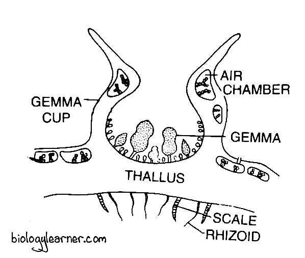
Gemmae Formation
The most common method of vegetative reproduction in Marchantia is the formation of special multicellular vegetative reproductive bodies called gemmae (singular: gemma). The gemmae are produced in large numbers in gemma cups.
Gamma cups are shallow, crescent-shaped structures present on the dorsal surface of the thallus in the midrib region. They are about 2 mm in diameter and 3 mm in height with smooth, spiny or fimbriate margins.
V. S. passing through the gemma cup shows two well-differentiated regions: the upper photosynthetic region and the inner storage region. The structure of both the photosynthetic and storage regions is similar to that of the thallus.
Intermingled with the gemmae in the cupule are many club-shaped mucilage hairs which secrete mucilage.
They absorb water, swell, and cause the gemmae to get detached, thereby helping in the dissemination of gemmae from the gemma cup.
Many small stalked gemmae develop on the bottom of the gemma cup.
Structure of Gemmae
Gemmae are small, discoid, biconvex (thick in the centre and thin at the apex) multicellular structures. They are autotrophic and green in colour.
Each gemma consists of a short, delicate, single-celled stalk. The stalk attaches it vertically to the bottom of the gemma cup.
Gemma is composed of many parenchymatous cells, oil cells, and rhizoidal cells. It is notched on two sides, in which lies the growing apex.
All the cells of gemma contain chloroplasts, except oil cells and rhizoidal cells. Oil cells are situated in the margins and contain oil bodies instead of chloroplasts. Rhizoidal cells are colourless, large in size, and present in the central region of the ventral side. These rhizoidal cells give rise to rhizoids on germination.
Dissemination of Gemmae
The mucilage hairs associated with gemmae help in the dissemination of gammae. They secrete mucilage on the absorption of water.
The mucilage swells up and presses the gemmae to disseminate from the stalks in the gemma cup. The gemmae are dispersed over long distances from the parent thallus by water currents.
Germination of Gemmae
The detached gemmae start germinating after falling on a suitable substratum. The surface of the gemma which comes in contact with the soil becomes the ventral surface.
Some colourless superficial cells are larger than the neighbouring cells and have dense and granular cytoplasm. These cells are called rhizoidal cells. The rhizoidal cells on the ventral surface of the gemma develop rhizoids.
Simultaneously, the apical cells present in the two lateral notches develop two thalli of new independent plants in the opposite direction. As a result, a single gemma gives rise to two thalli.
The gammae that develop on the male thallus form male plants, and those on the female thallus produce female plants.
Development of Gemmae
The gemma develops from a single superficial cell on the bottom of a gemma cup. The cell is papillate, known as the gamma initial.
The gamma initial undergoes a transverse division to form an upper cell and a lower stalk cell. The lower stalk cell forms the single-celled stalk.

The upper cell further divides by a transverse division and forms two cells. These two cells undergo similar divisions to form four cells. All these cells divide by vertical and horizontal division to develop a plate-like structure with two marginal notches, called the gamma.
Sexual Reproduction
Sexual reproduction in Marchantia is of the oogamous type. The sex organs are developed only under special environmental conditions such as long day length, high humidity, and low nitrogen content.
The sex organs are produced on special erect stalked modified lateral branches of the thallus called gametophores or receptacles. The gametophore bearing the male sex organ antheridium is called the antheridiophore, and the gametophore bearing the female sex organ archegonium is called the archegoniophore or carpocephalum.
Although the gametophores are actually direct prolongations of the prostrate vegetative thallus. This is clearly evident from the dorsiventral nature of the erect shoots with air chambers, airpores, photosynthetic tissues, scales, and rhizoids.
Marchantia is heterothallic (dioecious), i.e., antheridiophores and archegoniophores are developed on different plants. Species like M. palmata and M. polymorpha show abnormal receptacles bearing both archegonia and antheridia. Such receptacles are known as androgynophorous receptacles.
Archegonium develops on the undersurface and antheridium forms in the pit that opens outwards on the upper surface of the receptacles.
Archegonium develops on the undersurface and antheridium forms in the pit that opens outwards on the upper surface of the receptacle.
Antheridiophore
The antheridiophore is an erect modified branch that bears antheridia. It consists of a 1 to 3 cm long prismatic stalk and a disc-shaped receptacle at the apex.
The stalk is more or less cylindrical. The internal structure of the stalk shows air chambers and pores on its posterior side and two grooves or furrows on its anterior side. The two grooves run longitudinally the entire length of the stalk. Each of them contains rhizoids and scales.
The disc or receptacle is generally eight-lobed. In M. geminata, the disc is four-lobed. The internal structure of the peltate receptacle is similar to that of the vegetative thallus. It shows the upper epidermis with a number of air pores.
The pores open into underlying air chambers containing the photosynthetic filaments present in a horizontal row. Underneath the air chambers, there is a compact region of colourless parenchyma cells.

The lower epidermis contains scales and rhizoids.
Alternating with the air chamber on the upper surface of the receptacle are the flask-shaped cavities known as antheridial chambers. The antheridial chamber opens externally through a narrow pore called an ostiole.
The antheridia lie within the antheridial chambers. They are arranged in a radial row. A single antheridium in each chamber.
The antheridia are developed in an acropetal order, with older antheridia at the centre and younger ones at the margins.
Antheridium
The male reproductive organ of Marchantia is the Antheridium.
Development of Antheridium
- In Marchantia, the development of antheridium is similar to that of Riccia.
- The antheridium develops from a single superficial cell called the antheridial initial.
- The initial cell is situated on the dorsal surface of the receptacle, 2 or 3 cells back from the apical cell.
- The antheridial initial increases in size and divides transversely to form an upper outer cell and a lower basal cell.
- The basal cell remains embedded in the tissue of the thallus, undergoes a few divisions, and forms the embedded portion of the antheridial stalk.
- The outer cell divides by transverse divisions to form a filament of four cells.
- The upper two cells of the four-celled filament are primary antheridial cells, and the lower two cells are primary stalk cells.
- Primary stalk cells undergo a few further divisions to form the stalk of the antheridium.
- Primary antheridial cells divide by two successive vertical divisions at right angles to each other, forming two tiers of four cells each.
- Periclinal divisions now appear in both the tiers of four cells, and there is the formation of eight outer sterile jacket initials or wall initials and eight inner fertile primary androgonial cells.
- Jacket initials undergo several anticlinal divisions and form a single layer of the antheridial jacket.
- Primary androgonial cells undergo further divisions (transversely and vertically), resulting in the formation of a large number of small cubical androgonial cells.
- The last generation of the androgonial cells is known as sperm mother cells or spermatocytes (also known as the androcyte mother cells).
- The androcyte mother cell undergoes a diagonal mitotic division and forms two triangular cells called androcytes or spermatids.
- The androcytes, or spermatids, finally metamorphose into biflagellate antherozoids or sperm.

Structure of Antheridium
The mature antheridium is an elongated structure. It has a globular or pear-shaped body with a short multicellular stalk. The stalk attaches the antheridium to the base of the antheridial chamber.
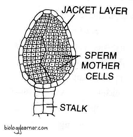
A sterile jacket layer encircles the body of the antheridium. The single-layered sterile jacket surrounds the mass of androcyte mother cells or sperm mother cells. Each androcyte mother cell divides to form two androcytes or spermatids. The androcytes metamorphose into antherozoids or sperm.
Antherozoids
The antherozoid is a unicellular, uninucleate, biflagellate, and rod-shaped or coiled structure.
A pair of whiplash flagella are present at the anterior end of the antherozoid. Both flagella are morphologically similar but differ in function. One flagellum serves for propulsion while the other is for rotation and for changes in direction.
Dehiscence of Antheridium
The mature antheridium dehisces with the help of water provided by rain or dew drops.
Water enters the antheridial chamber through the ostiole. Due to imbibition, the sterile apical cells of the antheridial jacket enlarge by absorbing water. They become softened and ultimately disintegrate.
Thus, the mature antheridium ruptures at its distal end. A large number of antherozoids come out just like a smoke column from the antheridial chamber onto the dorsal surface of the thallus. With the help of flagella, the antherozoids swim in the film of water.
Archegoniophore
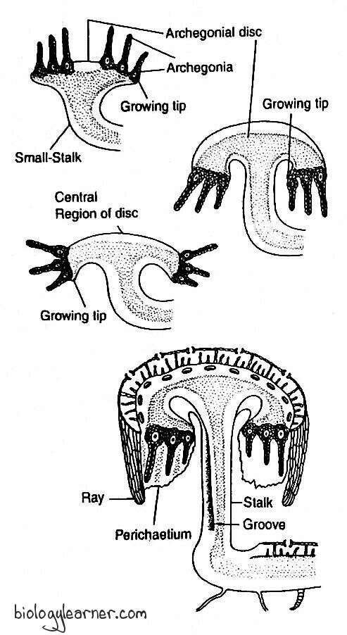
The archegoniophore is an erect modified branch that bears archegonia. It is also known as the carpocephalum.
Each archegoniophore arises at the apical notch of the thallus. It consists of a long stalk, slightly longer than the antheridiophore, and a disc-shaped receptacle at the apex. The stalk is maybe 5 to 7 cm long. The disc bears 8 rays or lobes (often 9 rays), and the growing apices of these rays bend downwards and inwards towards the stalk.

The archegonia develop on the upper surface of the young receptacles in an inverted position with their necks directed downwards. They are arranged in eight radial rows, corresponding to the eight lobes of the disc. There are 12-14 archegonia in each row.
The archegonia are developed in an acropetal order, i.e., the older archegonia at the centre and the younger ones at the margins.
Archegonium
The female reproductive organ of Marchantia is the archegonium.
Development of Archegonium
- Like the antheridium, the archegonium of Marchantia also develops from a single superficial cell on the dorsal surface of the young receptacle in an acropetal manner, called the archegonial initial.
- The superficial cell lies close to the apical cell.
- The conical-shaped archegonial initial becomes conspicuous and enlarges in size. It divides transversely into an upper primary archegonial cell (outer cell) and a lower primary stalk cell (basal cell).
- The primary stalk cell undergoes a few irregular divisions to form a short but distinct multicellular stalk of the archegonium.
- The primary archegonial cell enlarges and divides by three successive intercalary walls or periclinal vertical walls, resulting in the formation of three sterile peripheral initials and a fourth fertile median cell, the primary axial cell.
- The primary axial cell slightly overtops the peripheral initials.
- Each of the three peripheral initials divides by an anticlinal vertical division to form six jacket initials or envelope cells.
- In this way, the primary axial cell gets surrounded by jacket initials or envelope cells.
- Six jacket initials divide transversely into six jacket cells of the upper tier and six jacket cells of the lower tier.
- The upper tier is called neck initials, and it divides by repeated transverse divisions to form a tube-like neck.
- The neck of the archegonium is one cell thick.
- The cells of the neck are arranged in six vertical rows, and each row consists of six to nine cells.
- The lower tier is called venter initials, and it is also divided by repeated transverse divisions to form a single wall layer of the swollen venter.
- Simultaneously, the primary axial cell divides transversely and unequally to form a large lower central cell and a small upper primary cover cell.
- The central cell divides into a lower primary venter cell and an upper primary neck canal cell.
- The primary neck canal cell undergoes a series of transverse divisions and forms a row of about eight thin-walled neck canal cells.
- The primary ventral cell divides by an unequal transverse division to form an upper small venter canal cell and a lower large egg.
- The primary cover cell divides into two successive vertical divisions at right angles to each other, forming four cover cells which develop the mouth of the archegonium.

Structure of Archegonium
The mature archegonium is a short-stalked, pendulous, flask-shaped structure. The stalk attaches it to the surface of the receptacle.
The archegonium consists of an upper slender protruded neck and a basal swollen portion called the venter.
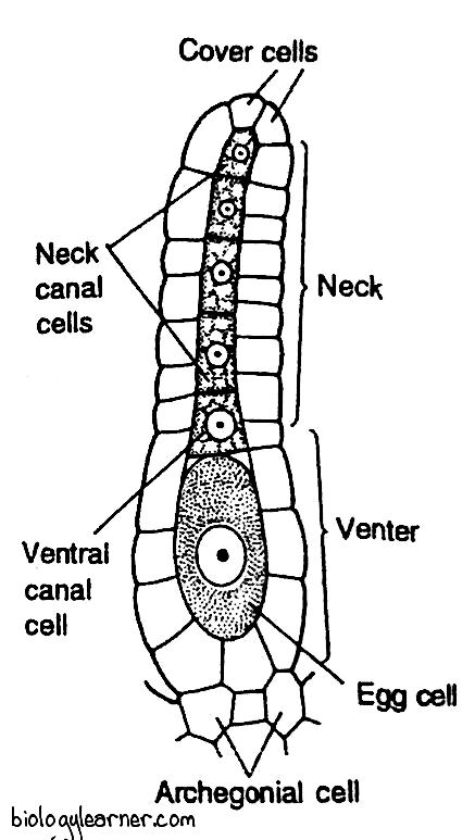
The long neck is a single-layered tube composed of six vertical rows of neck cells, characteristic of the Marchantiales order, and each row consists of six to nine cells. It encircles a narrow canal which consists of a row of eight neck canal cells. The apex of the neck is made up of four specialised and slightly large cover cells, or cap cells.
The venter is composed of a single-layered jacket or wall of sterile cells. The jacket encloses a small venter canal cell and a large naked egg.
Fertilization
Like other bryophytes, water is essential for fertilization in Marchantia. Due to its heterothallic (dioecious) nature, fertilization takes place only when the male and female plants grow near each other.
The process of fertilization occurs at a stage when the neck of the archegonium is in an upright position on the dorsal surface of the disc of the archegoniophore.
In the mature archegonium, the neck canal cells and the single ventral canal cell disintegrate to form a mucilaginous mass. The mucilage imbibes water, swells up, and comes out of the archegonial mouth by pushing the cover cells apart, forming a narrow passage called the neck canal. This mucilage mass contains chemical substances (chemical proteins or potassium salts) that attract the antherozoids.
The flattened, somewhat concave dorsal surface of the male disc functions as a shallow splash cup. Raindrops falling on the disc of the antheridiophore can splash the antherozoids up to two feet. This process is called the “splash cup mechanism.”
The antherozoids of the antherodiophore discs are splashed by raindrops and fall on the nearby female receptacle or may swim the whole way to reach the female receptacle.
A number of antherozoids enter the archegonial neck due to the chemotactic response and reach up to the egg. Ultimately, one antherozoid fertilises the egg to form a diploid zygote or oospore.
The gametophytic phase ends after the completion of fertilisation.
Post-Fertilization Changes (Inversion of Archegonia) in Marchantia
The following changes occur simultaneously after fertilization:
- The stalk of the archegoniophore elongates, becoming a couple of mm in length.
- The central sterile part of the disc enlarges massively. As a result, the marginal apical region of the disc containing archegonia is pushed downward and inward.
- The archegonia are now hanging towards the lower side of the disc with their neck portions pointing downwards.
- Between the rows of archegonia, long, green, finger-like projections called rays emerge from the margins of the disc. They give the female receptacle a stellate form as they radiate outward and curve downward. In M. polymorpha, the number of rays is nine.
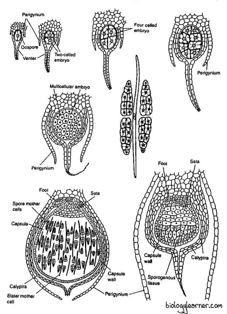
- The cells of the venter wall divide to form two to three-layered calyptra. It encloses the growing sporogonium.
- A ring of cells at the base of the venter divide and redivide to form a one-layered thick collar around the growing sporogonium, called perigynium or pseudoperianth.
- On either side of each row of archegonia, a one-celled thick, fringed sheath called perichaetium or involucre is developed. The involucre separates one row of archegonia from the other.
- Thus, the developing sporophyte is surrounded by three concentric protective sheaths of gametophytic origin; the calyptra, perigynium or pseudoperianth, and perichaetium or involucre. The major function of these sheaths is to provide protection against drought to young sporophytes.
Sporophyte of Marchantia
The sporophytic phase is the direct result of the sexual process and begins with the formation of the diploid zygote or oospore after fertilization.
The zygote, or oospore, is the first cell of the sporophyte.
Development of Sporophyte
- After fertilization, the diploid zygote immediately secretes a thin cellulose wall around itself. It enlarges in size and almost completely fills the cavity of the venter in the archegonium.
- The zygote divides first by a transverse division to form an outer epibasal cell and an inner hypobasal cell.
- The second division is at a right angle to the first (dividing vertically) and results in the formation of a four-celled embryo, or quadrant stage.
- Each quadrant is then divided by another vertical division at a right angle to the preceding one. As a result, the embryo develops into an eight-celled structure or octant stage.
- The four epibasal cells of the octant develop the capsule and upper part of the seta.
- The four hypobasal cells, undergo several divisions to form the lower part of the seta and the foot.
- Since the capsule forms the apex of the sporophyte and is developed from the epibasal cell, the type of embryogeny is described as exoscopic.
- The young eight-celled embryo now divides into all possible planes (divides irregularly) and forms a spherical mass.
- The lower cells divide to form a massive, bulbous foot of the sporophyte.
- The cells of the seta divide in one plane and form vertical rows of cells.
- When the embryo is about 12 or more cells, the cells in the upper region of the capsule now divide periclinally (parallel with the surface), resulting in the differentiation of an outer single layer of amphithecium and an inner mass of cells (multilayered) called endothecium.
- The cells of the amphithecium divide only by anticlinal walls to form a single-layered sterile jacket or capsule wall of the young sporophyte.
- The endothecium forms the archesporium, the first cell generation of sporogenous tissue.
- As the sporogonium advances towards maturity, the cells of the archesporium divide and re-divide several times to form a mass of sporogenous cells.
- The cells of the last cell generation of the sporogenous mass function as potential sporocytes or spore mother cells.
- The sporogenous cells in M. polymorpha undergo five successive divisions to form 32 spore mother cells, whereas, in M. domingensis, they divide only by three to four divisions to produce either eight or sixteen spore mother cells.
- Subsequently, half of the sporogenous cells become narrow and elongate to form the elater mother cells.
- The elater mother cells become elongated and form long, slender, sterile, diploid cells called elaters.
- Each elater possesses two spiral bands or thickenings on the surface of the wall and is pointed at both ends. They are hygroscopic and help in the dispersal of spores.
- Each of the spore mother cells is diploid and divides reductionally (meiotic division) to produce four haploid spores arranged tetrahedrally (spore tetrad).
- Later, the spores become free and remain surrounded by the capsule wall along with the elaters.
- In many species of Marchantia, such as M. polymorpha, the quadrant type of sporophyte development is quite common.
- But in a few species (e.g., M. chenopoda), a very rare type of embryo development occurs.
- Here the zygote is divided by two transverse divisions and forms the 3-celled filamentous embryo.
- In it, the hypobasal cell forms the foot and the middle seta, while the epibasal cell develops into a capsule of the sporophyte.
Structure of Sporophyte
The mature sporophyte of Marchantia is more or less an elongated structure. It can be differentiated into three distinct regions: the foot, seta, and capsule.
Foot
The basal portion of the sporophyte is the foot. It is multicellular, broad, bulbous or anchor-shaped.
The foot is embedded in the tissue of the archegoniophore (female gametophyte) and consists of parenchymatous cells. It helps in the anchorage of sporophytes and the absorption of water and nutrients from the gametophyte for the developing sporophyte.
Seta
The seta is a slightly enlarged stalk that connects the capsule with the foot. It contains numerous vertical rows of parenchymatous cells.
At maturity, due to several transverse divisions, the seta elongates rapidly and ruptures the calyptra. Ultimately, it pushes the capsule through the perigynium and perichaetium.
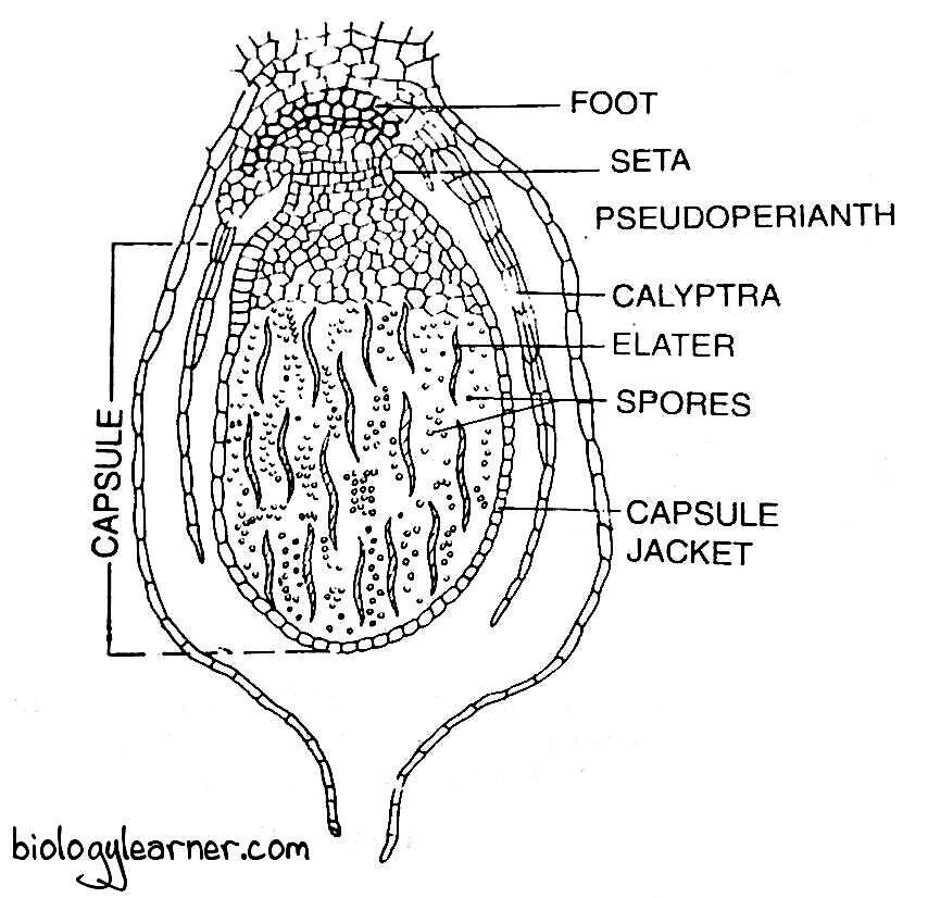
Capsule
The upper part of the sporophyte is the capsule. It is yellow-coloured, with an oval or spherical structure.
The capsule has a single-layered wall (sterile jacket wall) that encloses numerous spores and sterile elaters. It also contains calyptra, perigynium, and perichaetium, which collectively form a protective covering around the capsule.
Spore
The spores are haploid, uninucleate, and spherical in shape. They are very small, 0.012 to 0.03 mm in diameter.
Each spore remains surrounded by two wall layers: the outermost exospore or exine, and the innermost endospore or intine. The outermost exine is thick, smooth, and slightly reticulate, while the innermost intine is thin.
A tiny mass of granular cytoplasm is present inside the spore wall. It consists of a single nucleus and some reserve food material.
In a few species of Marchantia, such as M. torsana and M. caneiloba, the spores are tetrahedrally arranged. The perispore, an additional layer, is present outside the exine.
Dehiscence of the Capsule and Dispersal of Spores
As the sporophyte matures, the seta elongates rapidly and ruptures the calyptra. It pushes the mature capsule into the air through the perigynium and perichaetium.
The capsule wall (jacket) splits longitudinally from the apex to the middle by an indefinite number (four to six) of segments or valves. Due to the annular thickening in the cells of the capsule wall, the segments roll backwards, exposing the spores and elaters.
Hygroscopic movement in the elaters assists in the dispersal of spores.
The elaters are hygroscopic. They coil and uncoil with the changes in the moisture of the surrounding air. In dry weather, elaters lose water and become coiled. When the atmosphere is wet, they become uncoiled and cause the jerking action.
Due to the jerking action, the spore mass loosens. Finally, the spores are thrown into the air and dispersed by air currents.
Germination of Spores and Formation of Young Gametophyte
The spore is the first cell of the gametophytic generation. Each spore germinates immediately upon falling on a suitable moist substratum. They remain viable for about a year.
Under favourable conditions, the spores absorb moisture from the substratum and increase considerably in size. The chloroplasts now reappear.
Subsequently, the spore divides by transverse division and forms two unequal cells. The smaller cell is achlorophyllous and extends to form the first rhizoid. While the larger cell undergoes irregular divisions to form a six to eight-cell germ filament or protonema.
The contents of the cells move to the apex. The apex is cut off from the rest of the sporeling by a transverse division to form a small distal cell, which is wedge-shaped with two cutting faces. Now it functions as an apical cell.
The apical cell divides repeatedly to form a new Marchantia thallus.
Due to the heterothallic (dioecious) nature of Marchantia, the two spores of a spore tetrad develop into male thalli and the other two spores develop into female thalli.
