Riccia is the most widely distributed genus in the family Ricciaceae of the order Marchantiales. They commonly grow in moist soil, particularly during and after rain.
In Riccia, the gametophytic plant body is the dominant phase of the life cycle.

External Structure of Riccia
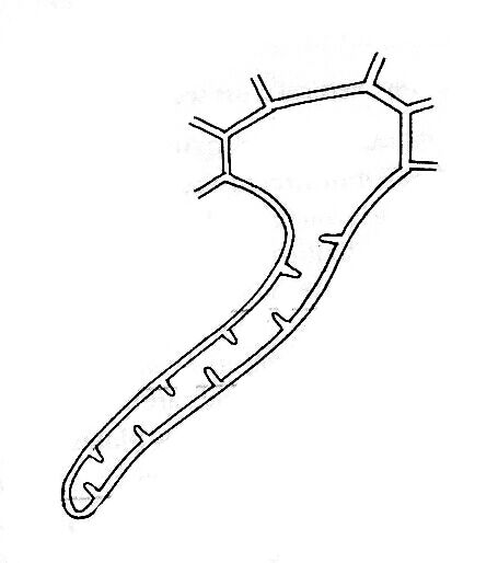
The plant body of Riccia is gametophytic. The gametophyte is a flat, dichotomously branched, dorsiventral prostrate, ribbon-like, green, fleshy thallus. Each branch of the thallus is linear to wedge-shaped or obcordate.
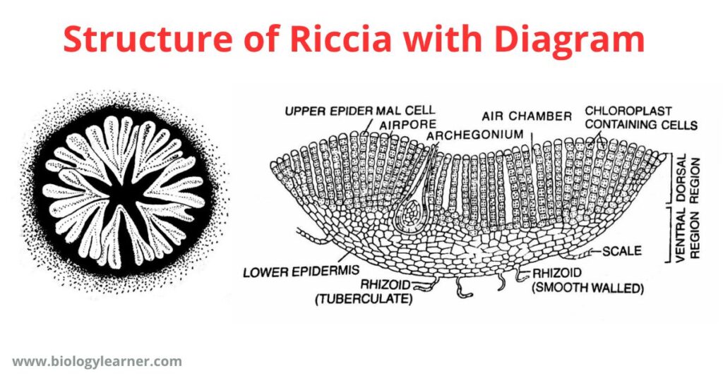
In terrestrial habits, the plants usually occur in a rosette form or a circular patch due to the presence of a number of dichotomous branches that grow together in one place.
The thallus is 5-7 mm long and 1.2 mm wide in all terrestrial species, while in Riccia fluitans, it is 30-50 mm long and 1-2 mm broad.
Dorsal Surface
The dorsal surface of the Riccia thallus is usually green in color, each branch having a thick midrib in the sagittal axis.
The midrib is traversed by a conspicuous median longitudinal furrow or groove which ends in a depression at the apical region of the thallus, forming an apical notch. The growing point is situated at the tip of the notch.
The main function of the mid-dorsal furrow is to retain the water required for fertilization.

Ventral Surface
The ventral surface of the Riccia thallus bears numerous scales and rhizoids.
Scales or Hairs
- Scales are multicellular, one-cell thick, and violet-colored structures that increase the surface area for absorption.
- The color of the scale is due to the pigment dissolving in the cell sap. The scales are more crowded near the apex and overlap each other at the growing point.
- Each scale splits up into two in the mature portion of the thallus and forms two rows of scales along the two margins of the thallus.
- They are ephemeral (short-lived) in hygrophilous species but persistent and leafy in xerophytic species. In Riccia crystallina, they are inconspicuous and absent.
Rhizoids
- The rhizoids are unicellular, elongated, unbranched, tubular hair-like structures present on the ventral surface of the thallus.
- They appear as prolongations of the lower epidermal cells.
- Mature rhizoids are devoid of protoplasm and are absent in aquatic forms (e.g., Riccia fluitans, R. natans).
- The main functions of rhizoids are to attach the thallus to the substratum and to absorb water and mineral nutrients from the soil.
- They are analogous to the roots of higher plants.
Rhizoids are of two types: smooth-walled rhizoids and tuberculate rhizoids.
- Smooth-walled rhizoids: The smooth-walled rhizoids are simple, elongated, and slightly wider. They contain a smooth inner wall with colorless contents.

- Tuberculate or pegged rhizoids: Tuberculate rhizoids are thinner and have peg-like or plate-like projections in the cell lumen.

Read also- Riccia: Distribution, Structure, Reproduction
Internal Structure of Riccia
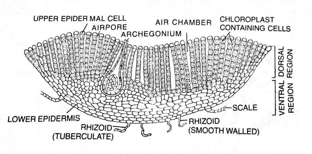
In a vertical cross-section, the thallus shows an internal differentiation of tissues distinctly organized into two horizontal regions, an upper photosynthetic or assimilatory region and a lower storage region.
Photosynthetic Region
- The upper dorsal region, i. e., the photosynthetic region, is composed of chlorophyll-bearing cells arranged in vertical rows.
- The chlorophyllous cells are separated by narrow longitudinal vertical air canals called air chambers.
- Each air chamber is generally surrounded by four vertical rows of cells (e.g., Riccia glauca), sometimes eight rows are also present (e.g, R. vesiculosa).
- The air chamber communicates with the external atmosphere through an air pore.
- Each air pore is a simple intracellular space bounded by 4 to 8 colorless enlarged terminal cells of the vertical rows which form a loose discontinuous one-celled thick upper epidermis.
- Air pores help in the gaseous exchange.

Storage Region
- The lower storage region of the thallus is composed of colorless, compactly arranged parenchymatous cells and lies below the photosynthetic region.
- The cells of the storage region (parenchymatous cells) are thin-walled, without intercellular spaces. They generally lack chlorophyll and contain starch granules as reserve food.
- The lower surface of the tissue contains small cells that are compactly arranged, forming a single layer of lower epidermis.
- Some cells of the lower epidermis extend to form the multicellular, one-celled thick scales and the unicellular tubular and smooth rhizoids.
