Marchantia is the most prominent genus in the family Marchantiaceae of the order Marchantiales. They prefer to grow in moist and shady places like wet open woodlands, banks of streams, wood rocks, or on shaded stub rocks.
A few species grow (e.g., Marchantia polymorpha) as the pioneers in burnt forest soil.
In Marchantia, the gametophytic plant body is the dominant phase of the life cycle, like that of Riccia.

External Structure of Marchantia
The plant body of Marchantia is gametophytic. The gametophyte is a flat, prostrate, dorsiventral, dichotomously branched thallus.
Dorsal Surface
The dorsal surface of the Marchantia thallus is dark green, with each branch having a median, more or less conspicuous midrib. The midrib in each branch of the thallus is marked on the dorsal surface by a shallow groove and on the ventral surface by a low ridge.
The dorsal surface of the thallus has a number of regular polygonal or rhomboidal areas called areolae. These polygonal areas demarcate the outline of the underlying air chamber. Each polygonal area has a pore at the center called an air pore.
Each branch of the thallus has a growing point situated at the apex of a terminal groove called an apical notch.
The dorsal surface of the mature thallus bears the vegetative and sexual reproductive structures. The vegetative reproductive structures are gamma cups, which develop along with the midrib. The gamma cups are cup-like or crescent-shaped structures with spiny or fimbriate margins.

Sexual reproductive structures (sex organs) are borne on special erect branches called gametophores or gamctangiophores. These branches are umbrella-shaped and arise from the distal end of the thallus at the growing point (apical notch).
The gametophores bearing antheridia (male sex organs) are called antheridiophores, and those bearing archegonia (female sex organs) are called archegoniophores. Marchantia is a heterothallic (dioecious) plant. Therefore, a thallus bears either antheridiophores or archegoniophores.
Ventral Surface
The ventral surface of the Marchantia thallus bears numerous scales and rhizoids along the midrib.
Scales
- The scales are multicellular, one-cell thick, and usually violet-colored structures that increase the surface area for absorption.
- The color of the scale is due to the presence of anthocyanin pigments.
- They are arranged in 3–4 rows on both sides of the midrib.
- Scales mainly protect the growing point and retain water by capillary action.
Scales are of two types: appendiculate and ligulate.
- Appendiculate scales: The appendiculate scales are larger and more elaborate due to the presence of an apical sub-rounded appendage. They form the inner row of the scales, close to the midrib.
- Simple or ligulate scales: The ligulate scales are relatively smaller than the appendiculate scales and without appendages. They form the outer or marginal row.
Rhizoids
- The rhizoids are unicellular, branched, colorless structures present between the scales on the ventral surface of the thallus.
- They develop as prolongations of the lower epidermal cells.
- The main functions of rhizoids are to attach the thallus to the substratum and to absorb water and mineral nutrients from the soil.
Rhizoids are of two types: smooth-walled rhizoids and tuberculate rhizoids.
- Smooth-walled rhizoids: The smooth-walled rhizoids are simple, elongated, and slightly wider. They contain a smooth inner wall with colorless contents.
- Tuberculate or pegged rhizoids: Tuberculate rhizoids are thinner and have peg-like or plate-like projections in the cell lumen. They appear like circular dots on the surface view.
Read also- Marchantia: Distribution, Structure, Reproduction
Internal Structure of Marchantia
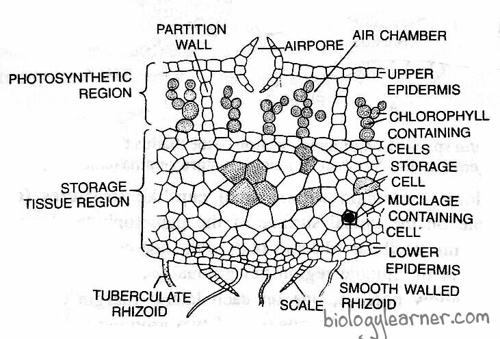
In a vertical cross-section, the Marchantia thallus shows an internal differentiation of tissues distinctly organized into three regions: the epidermal region, the photosynthetic region, and the storage region.

Epidermal Region
- The epidermal region is composed of a well-defined upper and lower epidermis.
- The epidermis is formed of thin-walled, quadrate cells containing a few chloroplasts.
- There are a number of air chambers that are present in a single horizontal layer just below the upper epidermis.
- The air chambers are connected with the outside atmosphere by a barrel-shaped air pore.
- The air pore is surrounded by four to eight superimposed tiers of concentric rings, each tier containing three to four cells. Air pores help in the gaseous exchange.
- The lower tier consists of four cells that project into the pore, and the opening of the pore looks star-like in the surface view. The lowermost ventral layer is the lower epidermis, which develops scales and rhizoids.
Photosynthetic Region
- The photosynthetic region lies below the upper epidermis.
- A single-layered partition of cells (partition walls) containing chloroplasts separates the air chamber from the others.
- The partition walls are two to four cells in height.
- Many simple or branched photosynthetic filaments arise from the base of each air chamber. These filaments are formed of chloroplasts bearing cells in a horizontal row.
Storage Region
- The storage region lies below the photosynthetic region.
- It is composed of several layers of compactly arranged polygonal parenchymatous cells.
- The parenchymatous cells are thin-walled and lack intercellular spaces and chloroplasts.
- The cells of the storage region generally contain starch grains or protein granules. Large oil bodies or mucilage are also found in some cells.
- The cells of the midrib region are elongated, showing reticulated thickening.
