Sclerenchyma is a type of simple permanent tissue that provides structural support to the plant. It consists of thick-walled, lignified sclerenchyma cells. The cells mostly lack protoplasm at maturity and become dead.
Mettenius first coined the term sclerenchyma. It is derived from the Greek words sclerous and enchyma. Sclerous means hard.
Definition of Sclerenchyma Cells
Sclerenchyma cells are heavily lignified, thick-walled cells that mainly provide mechanical stiffness and strength to plants.
Distribution
- Sclerenchyma cells are usually present in the cortex, pericycle, pith, xylem, and phloem.
- They also occur in the pericarp of fruits (e.g., Psidium, Pyrus) and thick testa of the seeds (e.g., Pisum, Vigna, Lathyrus).
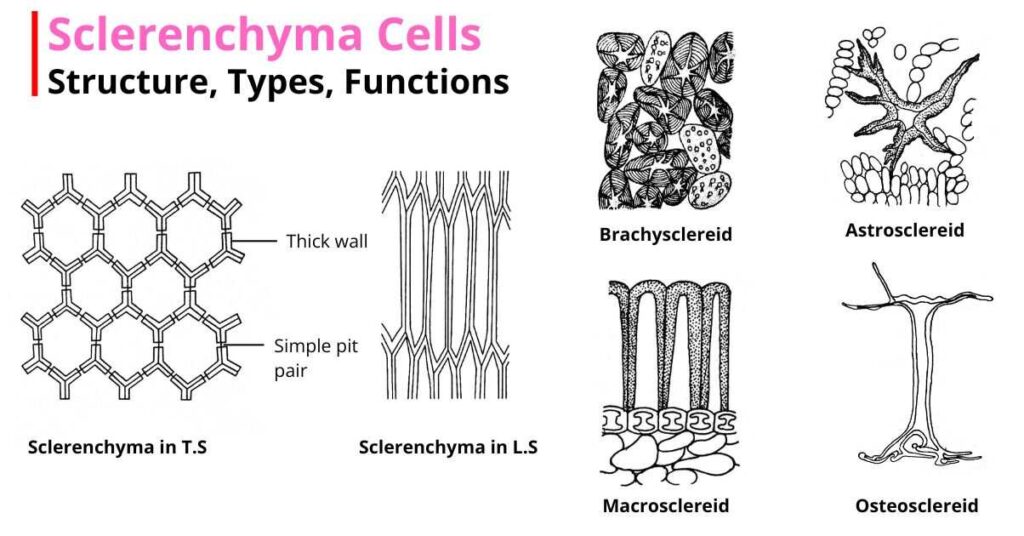
Structure of Sclerenchyma cells
- The sclerenchyma cells are various shapes and sizes. They may be niddle-like, elongated, spherical, or star-shaped.
- The cell wall is uniformly thick and often strongly lignified. Thus, the walls of sclerenchyma cells are more rigid in comparison to the parenchyma cells and the collenchyma cells.
- There are also cutin and suberin, in the cell walls, making the walls waterproof.
- Presence of simple pits in the walls.
- The mature cells are dead and do not possess protoplasm.
- Intercellular spaces are absent, as the cells are closely packed.
Types of Sclerenchyma Cells
Sclerenchyma cells show great variation in their form, structure, origin, and development. Most commonly, they are categorized into two groups:
- Fibres and
- Sclereids or sclerotic cells.
Fibres
- Fibres are elongated sclerenchyma cells with pointed or tapered ends.
- Sometimes, cells with branched ends may also be found.
- Fibres are much longer than their breadth. They are the longest cells in plants. In Boehmeria nivea, they are up to 55 cm long.
- The cell walls are hard, uniformly thickened, and often heavily lignified.
- The central cavity or lumen is almost blocked due to so much thickening of the secondary wall.
- The pits are small, round, or slit-like.
- The cells gradually lose their protoplasm and become dead and empty at maturity.
- Fibres may occur in patches with overlapped or interlocked ends.
- They are found in the cortex and pericycle of many dicots, xylem, and phloem tissues.
According to their position in the plant body, fibres are of two types:
1. Xylary Fibres or Wood Fibres
The fibres associated with xylem are called xylary fibres.
These are of two types: libriform fibres and fibre-tracheids.
- Libriform Fibres are the narrow fibres and have highly lignified, thickened secondary walls with reduced, simple pits. They look like phloem fibres.
- Fibre-tracheids are intermediate forms between fibres and tracheids. They have relatively thin walls with much-bordered pits and are often regarded as reduced tracheids.
2. Extraxylary Fibres or Bast Fibres
The fibres occurring outside the xylem are called extraxylary fibres or bast fibres. They are usually long and spindle-shaped, with blunt ends. The walls are highly thick and either lignified or non-lignified. Pits may be simple or slightly bordered.
Bast fibres are of the following types on the basis of their topography:
- Phloem fibres that occur in the primary and secondary phloems.
- Cortical fibres that are found in the cortex.
- Perivascular fibres that are present in the peripheral part of the vascular cylinder inside the innermost cortex.
Sclereids or Sclerotic Cells
- Sclereids are modified sclerenchyma cells, usually isodiametric or irregular in shape.
- They occur either singly or in groups in the plant body.
- The wall is thick and lignified. The thickness may vary in different cells. As the cell walls are very hard, sclereids are also called stone cells.
- Many simple pits with round apertures are present in the cell walls. Sometimes, bordered pits may also be found.
- Cells usually have a narrow lumen.
- Mature sclereids are dead cells, but it has been observed that in some cases they can retain living protoplasm.
- Sclerotic cells are mainly found in association with xylem and phloem tissues.
According to the size, shape and the nature of the cell walls, sclereids are classified into the following five groups (Bloch, 1946):
1. Brachysclereids
- These are parenchyma-like cells. The cells are short and isodiametric.
- Due to the gritty texture of the pulp, these cells are also known as grit cells.
- Brachysclereids are abundantly found in the cortex, pith, and phloem of stems and in the pulp of some fruits like Pyrus and Psidium.
2. Macrosclereids
- These are elongated, rod-like, or columnar sclereids.
- They form the palisade-like epidermis of the seed coats of many leguminous plants (e.g., Pisum, Phaseolus).
- They are also found in the leaves of xerophytes.
3. Osteosclereids
- These are bone-shaped cells.
- The cells are columnar but enlarged at their ends.
- They are found in the hypodermal layer of the seeds and fruits of many dicots.
4. Asterosclereids
- In this type, cells are star-shaped.
- The cells have irregularly branched arms, which give them a star-like or stellate appearance.
- Astrosclereids occur in the intercellular spaces of the leaves and stems of some hydrophytes (e.g., Nymphaea).
5. Trichosclereids
- These are hair-like, much elongated, and branched sclerotic cells.
- The branching can extend into the intercellular spaces.
- Trichosclereids are found in the leaves of Olea and the aerial roots of Monstera.
Functions of Sclerenchyma Cells
- Sclerenchyma cells mainly provide mechanical strength and rigidity to the plant.
- They protect the plant from stress and strain.
- Fibres obtained from jute, hemp, and flax have great commercial value.
