Restriction enzymes are the endonuclease enzymes that cut the double-stranded DNA at specific sites, resulting in fragments of DNA. So these enzymes are called molecular scissors. Restriction enzymes are present in bacteria and provide a type of defense mechanism (the restriction-modification system).
This type of endonucleases (restriction enzymes) recognize and cleave the specific DNA sequences called restriction sites, for example, EcoRI (isolated from E. coli) that recognizes and cleaves the sequence 5′-GAATTC-3′ to generate cohesive or sticky ends. Similarly, HindIII isolated from Haemophilus influenzae recognizes and cleaves the sequences 5′-AAGCTT-3′ to generate cohesive or sticky ends.
Enzyme activity is represented as IU (International Unit). One unit of a restriction enzyme is the amount of enzyme required to completely digest one microgram of lambda DNA (in a reaction volume of 50 μl) in one hour under optimal conditions of salt, pH, and temperature (about 37°C for most restriction enzymes).
Objective
Digestion of bacteriophage lambda (λ) DNA using a restriction enzyme.
Principle
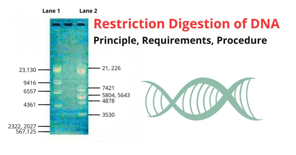
Phage lambda (λ) DNA is a linear double-stranded DNA containing 48502 base pairs (bp). Its DNA becomes circularised after release (inside the cell of E. coli) at a cohesive site called the COS site. It contains five recognition sites for EcoRI and seven recognition sites for HindIII. The complete digestion of lambda DNA with EcoRI results in 21, 226, 7421, 5804, 4878, and 3530 bp long six DNA fragments.
Similarly, complete digestion of lambda DNA with HindIII results in eight DNA fragments viz, 23, 130, 9416, 6557, 4361, 2322, 2027, 564, and 125 bp long fragments.
Requirements for Restriction Digestion of DNA
- Lambda DNA, restriction enzyme such as EcoRI or HindIII
- Assay buffer for the restriction enzyme, sterilized water, Tips, Eppendorf tubes, micropipettes
- Agarose gel, electrophoresis apparatus
- 5X TBE (Tris-Borate-EDTA) or 5X TAE (Tris-Acetate-EDTA)
Procedure of Restriction Digestion of DNA
- Always keep restriction enzyme (EcoRI or HindIII), substrate (lambda DNA), and assay buffer in an ice bucket.
- Take 2-5 μg of the lambda DNA as substrate in an Eppendorf tube and dissolve it in an appropriate volume of water.
- Add ul of about 10X assay buffer (available with the restriction enzyme) to the DNA in the Eppendorf tube, followed by the respective enzyme (5-12 units of EcoRI or 10-25 units of HindIII depending upon the amount of DNA used in the reaction step (previous step).
- Add sterilized water to make the final volume of the reaction mixture 20 μl. Centrifuge gently or mix by tapping with fingers.
- Incubate the reaction mixture for 1 hour at 37°C in a water bath or incubator.
- Prepare 1% agarose gel for loading and electrophoresis.
Preparation of 1% Agarose Gel and Set up of Electrophoresis
It follows the following steps:
- Dilute 50 X TAE or 5X TBE buffer with distilled water to get IX TAE or IX TBE.
- Pour 50 ml of IX-TAE or IX-TBE buffer into a 250 ml conical flask and add 0.5 g of agarose into it. Boil to get a clear solution and cool to warm a liquid (at 60°C).
- In the electrophoresis, set put the combs in such a way that they should be about 2 cm away from the cathode.
- Add ethidium bromide (10 mg /ml stock) to make a final concentration of 0.5 μg/ml of gel when the temperature of agarose gel is around 60°C.
- Gently pour the solution into the gel tank. Pour the agarose gel in such a way that it could be 0.5-0.9 cm thick and without air bubbles. Allow the gel to get solidified.
- After one hour stop the reaction by addition of 3.33 μl of 6X gel loading buffer to the Eppendorf tube. Label the vial as ‘A’ and put it on ice.
- As described in the second step, take the same amount of lambda DNA in another fresh 1.5 ml Eppendorf tube and mark it as ‘B’.
- On a 1% agarose gel, load samples A and B in separate wells. Note the order of the samples loaded in each well.
- Run it until the bromophenol blue dye has reached 3/4 of the gel (it takes about 1 hour). Observe under UV transilluminator.
Results
A single band is seen in the lane in which sample ‘B’ (lambda DNA) has been loaded. ‘A’ has been loaded and shows multiple bands. This reveals the cleavage of sample ‘B’ by the respective restriction enzymes.

The exact number and size of the bands obtained depend on the restriction enzymes used for digesting the lambda DNA (sample ‘B’). As explained earlier, 6 DNA fragments ( 21, 226, 7421, 5804, 4878, and 3530 bp) are observed if EcoRI is used and 8 DNA fragments (23, 130, 9416, 6557, 4361, 2322, 2027, 564, and 125 bp) are observed after using HindIII.
