Jungermanniales is the largest order of class Hepaticopsida in the division Bryophyta.
Characteristics of Jungermanniales
- This order contains 244 genera and more than 9000 species.
- Members of Jungermanniales are known as leafy liverworts. Because the gametophytes of this order are mostly foliose type and they are differentiated into stem and leaves.
- In some genera, gametophytes are thallose types like those of Marchantiales.
- Intermediate forms between thallose and leafy types are also noted among some members of this order.
- But the presence of any air chamber or any internal tissue differentiation like the gametophytes of Marchantiales is absent.
- The gametophytes bear only smooth-walled rhizoids.
- Scales are absent.
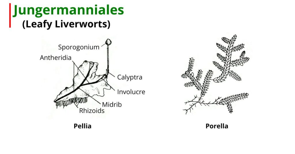
Structure of Gametophyte in Jungermanniales
The plant body is gametophytic. The gametophytes are thalloid, except Fossombronia.
External Morphology
- The plants are thalloid in which midrib is present (Pellia) but maybe absent (Riccordia).
- In Fossobronia, the plant body is erect and foliose, which is divisible in stem and leaves.
- In Pellia, the gametophytic plant body is thin, dorsiventrally flattened, prostrate, and dark bright green in color.
- Leaves are arranged in lateral margins, and a clear differentiation is observed in Fossombronia.
- Scales and rhizoids are absent in most plants. But in Pellia, numerous simple, smooth-walled, unicellular rhizoids are present irregularly on the mid-ventral surface of the thallus.
Internal Morphology
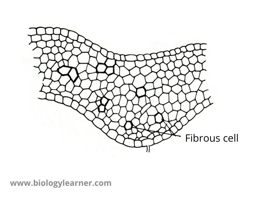
- The internal structure of the thallus is very simple. No tissue differentiation is observed in the thallus.
- Simple, thin-walled chlorenchymatous tissues are present between the upper and lower epidermis. From the lower epidermis rhizoids arise in Pellia.
- A single large apical cell situated in the apical notch brings out the growth of the thallus.
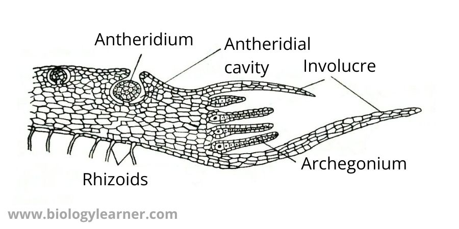
Sexual Reproductive Structures of Jungermanniales
- In some species, sex organs are developed at specific branches called antheridial and archegonial branches.
- Fossombronia is heterothallic, hence the male and female organs develop at separate thallus.
- In Pellia, the thallus may be monoecious (Pellia epiphylla) or maybe dioecious (P. endivaefolia). Sex organs (antheridium and archegonium) are borne on the thallus.
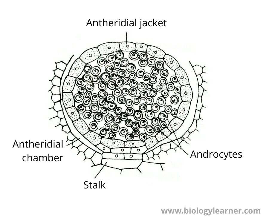
Antheridium
- Antheridium is the male sex organ.
- In antheridium, any cell behaves like a superficial cell and divides by transverse division to form an upper cell and a basal cell.
- Generally, the basal cell is inactivated and the upper cell divides to form a stalk cell and a primary antheridial cell.
- Stalk cell divides to form 2-celled stalk, while antheridial cell divided by transverse division and oblique division through which peripheral cells and central cells are formed.
- Peripheral cells are divided and a one-celled thick jacket is developed, while central cells divide to form antherozoid mother cells.
- Antherozoid mother cells produce antherozoids which are spiral, biflagellate, and uninucleate.
- In such a way, mature antheridium is made up of a 2 celled stalk, and a club-shaped structure.
Archegonium
- Archegonium is the female sex organ developed at the female thallus.
- In Pellia, The group of archegonia is protected by an involucre (an outgrowth of the thallus tissue developing from the base of the archegonium).
- In archegonium, any cell behaves like a superficial cell which is divided by transverse division to form an upper cell and a basal cell.
- Basal cell forms stalk. While the upper cell divides by oblique division to form peripheral cells and central cells.
- Peripheral cells continue to divide to form a jacket and the central cell divide to form the primary cover cell and central cell.
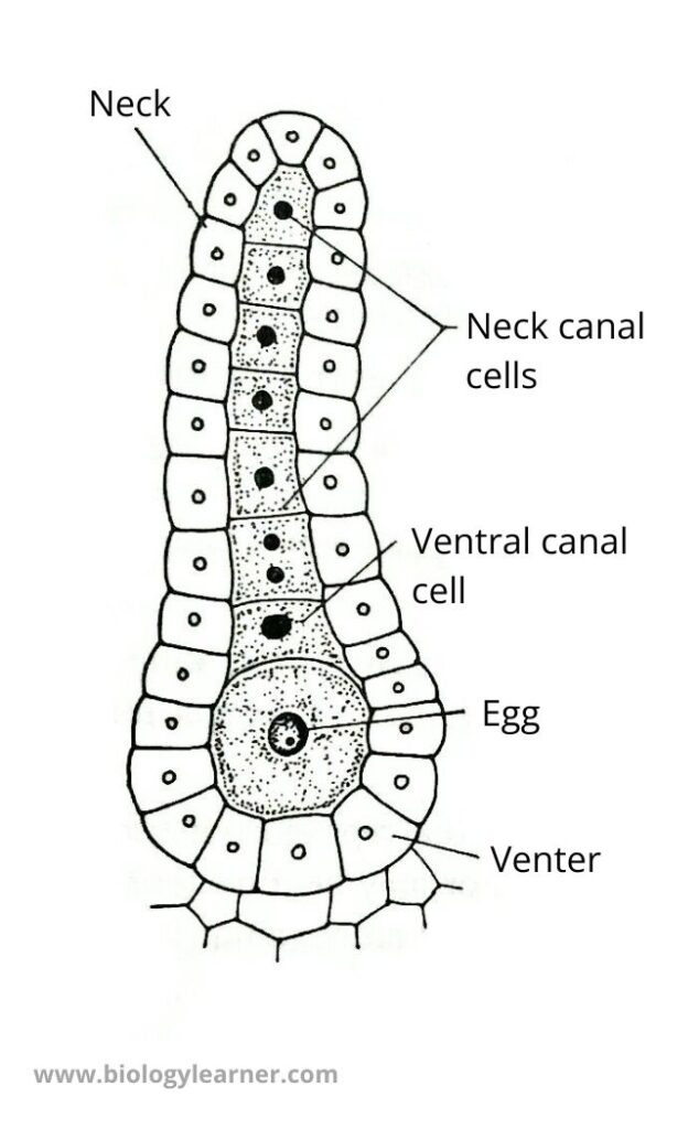
- The primary cover cell produces 4 cover cells and the central cell forms a primary neck initial and a primary venter initial.
- The Neck initial divides to form a neck canal cell and the primary venter initial divides to form one ventral canal cell and an egg.
- In such a way, mature archegonium consists of 4 cover cells, 6-8 neck canal cells, one ventral canal cell, and an egg.
Fertilization
- When archegonium matures and the antherozoid is released towards the archegonium, neck canal cells and ventral canal cells dissolved and water enters in neck portion.
- From the neck of the archegonium, some salts or proteins are excreted which stimulates the antherozoids to move in that direction.
- Then the antherozoid enters the archegonium male nucleus fused with an egg to form a zygote.
Development of Sporophyte
- A zygote is the first cell of the sporophyte, immediately after fertilization, the zygote increases in size.
- The zygote is divided by transverse division to form epibasal cells and hypobasal cells.
- Hypobasal cell is inactivated, while epibasal cell divides through 3 transverse, one longitudinal, and one vertical division.
- In this process, 4 tiers of 4-4 cells are formed.
- Upper 2-tiers form capsule.
- In upper tiers, periclinal division takes place through which outer amphethecium and inner endothecium are formed.
- Amphethecium develops in the outer jacket, while outer endothecium develops in the inner jacket.
- Inner endothecium changed into archesporial tissue, in which some are fertile and some are sterile.
- Fertile cells undergo meiosis to form spore-tetrad but sterile cells are present either at the lower portion (Pellia) or at the upper portion (Riccardia).
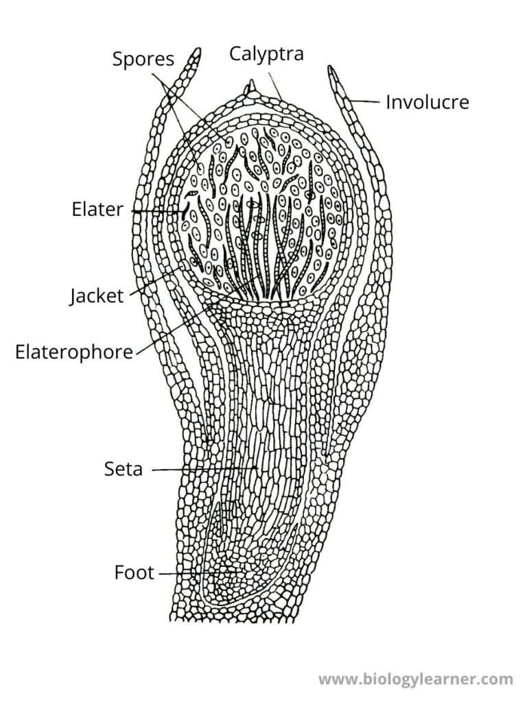
- These cells are elongated and form elaterophores. The elaterophores produce elaters.
- The elaters are spindle-shaped elongated structures provided with 2-3 spiral thickening bands.
Structure of Sporophyte
- The sporophyte consists of the foot, seta, and capsule.
- The foot is a small bulbous type and the seta is very short.
- The capsule is made up of 2 layers of thick jacket and inner side elaterophores.
- In elaterophores, elaters and spore-tetrads are present.
Dehiscence of Capsule
- When the capsule is matured then the upper jacket is dried away while the lower jacket is moist, so due to contraction and expansion capsule is opened into 4 slits.
- Its opening is similar to a flower opening.
Examples of Jungermanniales
Some common genera of Order Jungermanniales is Pellia, Porella, Fossombronia, Riccardia, etc.
Classification of Jungermanniales
Smith (1938) has divided Order Jungermanniales into two sub-orders:
- Metzgerineae
- Jungermannineae
