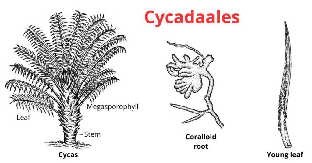
Cycadales is a gymnospermic order. Members of this order are mostly inhabitants of tropical and subtropical regions. It includes both living and fossil genera.
They originated in the Mesozoic era and continued up to the Jurassic and Cretaceous periods. Only ten genera are visible now. These genera are found in Australia, Central America, South Africa, and Eastern Asia. Cycadales are often with stout trunks. Stems are large pith, scanty wood, and thick cortex. Plants are woody, branched or unbranched, and dioecious.
This order includes only one family Cycadaceae (Pilger and Melchior, 1954). Remarkable characteristics present in their body show that they are the intermediate plants in between the ferns and typical gymnosperms. These are known as Cycad ferns, being the earliest seed-producing plants, showing fern-like appearances.
Distribution of Cycadales
- The members of Cycadales are mostly restricted to tropical and sub-tropical regions of the world.
- It includes 16 living genera and 1 fossil genus.
- They are mostly found in Australia, South Japan, Western China, India, Mexico, South America, and Cuba.
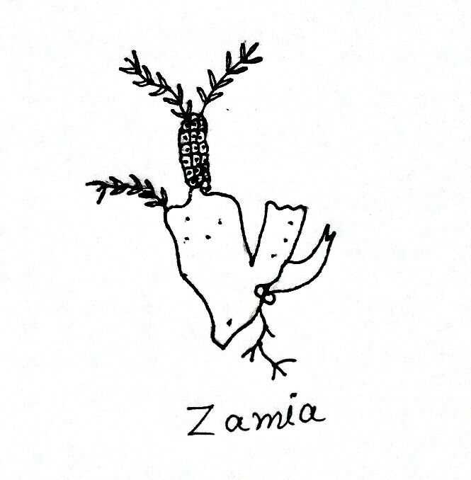
- Examples: Some common genera of Cycadales are Zamia, Lepidozamia, Bowenia, Dioon, Cycas, etc.
Morphology of Cycadales
Morphology of Cycadales is two types: External morphology (external structure) and Internal morphology (anatomical structure).
External Morphology
- These plants appear palm-like. The stem is thick cylindrical and unbranched. It is 10-18 meters in length like Dioon spinulosum.
- The stem of Bowenia is spherical, tuberous, and unbranched leaves are spirally arranged and are pinnate and bipinnate. Venation is open and dichotomous.
- In Cycas and Dioon leaves are dimorphic.
- Plants take about 1000 years to reach 6-7 feet in height.
Internal Morphology
Stem Anatomy
- The internal structure of the stem includes the peripheral cortex and large pith. Cortex and Pith have large mucilage canals.
- Endodermis and pericycle can be differentiated.
- Vascular bundles are arranged in a ring and conjoint collateral, open and endarch.
- Leaf traces are present.When branches develop, leaf traces arrange themselves in an irregular manner, which is an important characteristic of this order. These are also arranged in horseshoe shapes.
- Traces are diploxylic which has centripetal and centrifugal xylem.
- The secondary xylem has fewer amounts and is interrupted by multiseriate medullary rays.
- The wood is sort and is known as monoxylic wood.
- Various genera have single cambium, which is active throughout the life in plants. But in Cycas, Macrozamia, and Encephalartos, wood is polyxylic.
- Tracheids of Zamia and Stangeria have scalariform pits, but other genera have multiseriate-bordered pits.
- Medullary rays in Dioon are one cell thick and while in Cycas it is 6-7 cells thick.
Leaf Anatomy
- In leaves, sunken stomata are present, which are haplochellic.
- Vascular bundles are diploxylic and arranged centripetally in a triangular fashion. It has a single protoxylem group.
- The centrifugal xylem is present and is separated from the parenchyma (secondary xylem).
- The hypodermis is sclerenchymatous.
- Mesophyll tissue is divided into palisade and spongy tissues.
Internal Structure of Root
- Roots are very long and tetrahedral.
- Secondary growth is irregular.
- Roots are apogeotropic, so their branches shift from soil to the upper ground surface.
- The root tip is destroyed by the microbe’s enteric roots and is thus known as the coralloid root.
- Coralloid roots have one or rarely more than one-layered thick algal zone (Anabaena is present) in the cortex.
Reproduction of Cycadales
These plants are dioecious as the male and female flowers (reproductive structures) occur on different plants. Flowers are unisexual and simple, male flowers are represented by microsporophylls (stamens) and females by megasporophylls (carpels).
Female Strobilus
- Cycas megasporophylls are spirally arranged.
- The size of female cones (strobili) are vary 2-90 cm in different genera of Cyadales (Macrozamia denisonii – 70 cm, Dioon spinulosum – 50 cm, Dioon edule – 30 cm, Microcycas – 94 cm, and Zamia pigmea – 02 cm).
- Cycas has the largest female cone.
- In Cycas, there are many megasporophylls. Its upper part has a leaflet in which ovules are present. Ovules are terminal.
- In Dioon, cones are broad and loose.
- In Zamia, sporophylls are reduced and cones are compact. The terminal part of Ceratozamia is depressed.
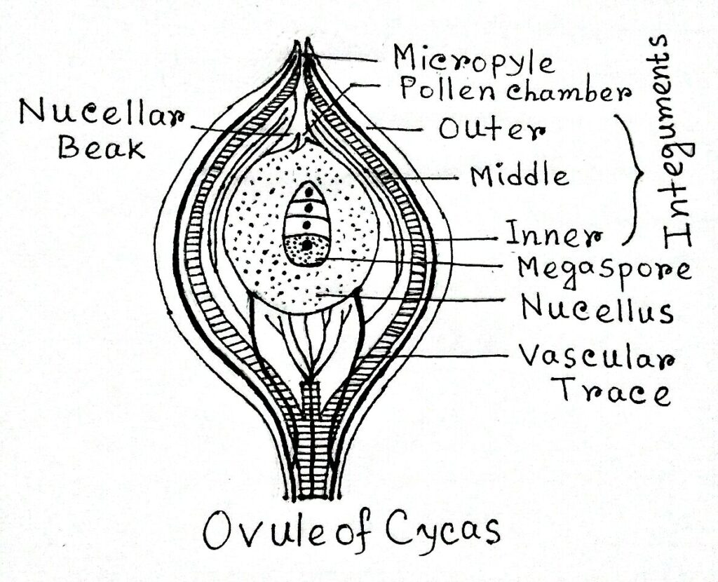
- Megasporangium is also called an ovule. It is unitegmic.
- The upper part has an integument, surrounded by a fleshy layer.
- The micropyle is present at the apex.
- Not much is known about the development of megasporangium.
- According to Smith, four megaspores develop in Zamia, four rows of spores are present in which the only one is functional for forming female gametophytes
Male Strobilus
- In sporophyll of male strobilus, leaves are absent.
- Cone is compact and the terminal cone is present.
- In Macrozamia denisonii, the largest cone was found which is 60-80 cm long.
- It is 40-45 cm long in Cycas and in Bowenia it is 5 cm long.
- Microsporophyll is arranged spirally in an acropetal manner.
- The shape of microsporophyll is variable.
- Microsporangium is abaxial and arranged radially in sori.
- Cycas has five sori and Zamia has single sori.
- Sporangia are formed of many cells and are thick-walled.
- The epidermis is unicellular and thick tapetum present which is one cell thick.
- Sporangium shows Eusporangiate type of development.
- In the center, sporogenous tissues are formed which in turn form spore mother cells.
- Spore mother cell divides by meiosis to form 4-microspores.
Gametophytes of Cycadales
Gametophytic generation begins with the formation of gametophytes. Then the fertilization occurs and The embryo is formed. In the end, seeds are germinated.
Female Gametophyte
- Megaspore is the first cell of the female gametophyte.
- During gametophyte formation, it increases in size and the adjacent cells form spongy tissues.
- First of all, free nuclear division occurs and many nuclei are formed. Nuclei shift toward the periphery due to the formation of the central vacuole.
- Now cell wall forms at the periphery zone which later moves towards the center.
- After that, a multicelled gametophyte is formed.The gametophyte is surrounded from all sides by 1-2 layered thick endosperm.
- During development few micropylar cells become enlarged, which are also known as archegonial initials.
- In Dioon edule, these initials are formed in November.
- It divides and forms a central cell. Along with the central cell, primary neck cells are also formed. Neck cells are two in number.
- The central cell gets enlarged its protoplasm gets vacuolated. It divides to form a ventral canal nucleus and an egg nucleus.
- Later, the ventral canal nucleus gets disappeared.
Male Gametophyte
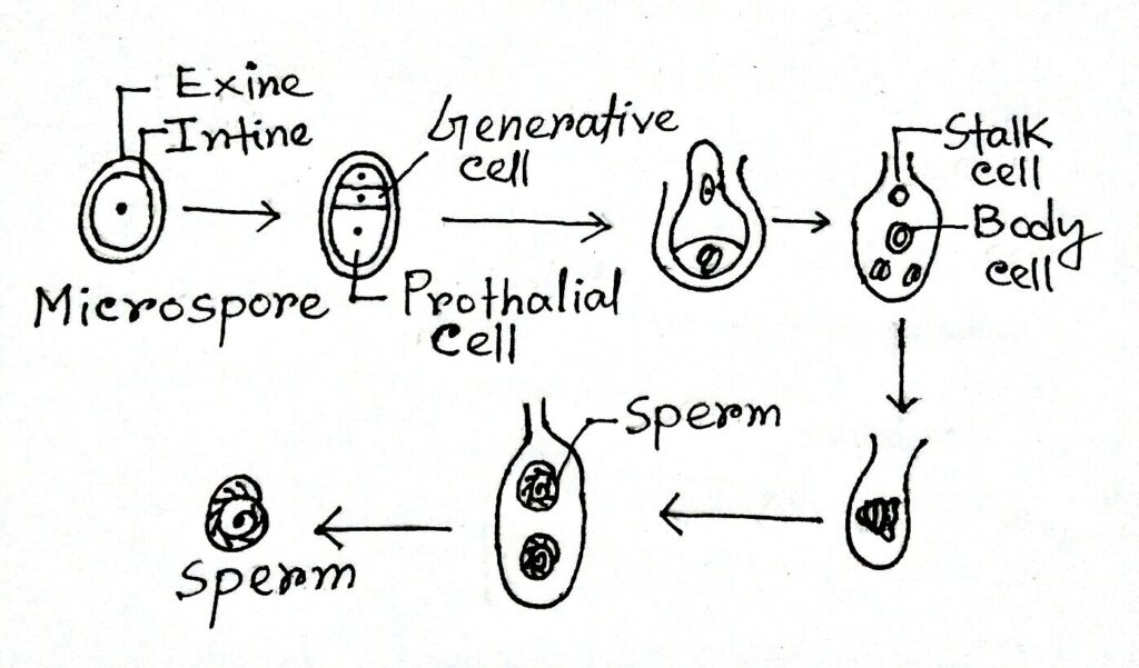
- Microspore is the first cell of the male gametophyte. It has exine and intine.
- The exine is thick and intine thin.
- Micospores are in microsporangium.
- Prothalial cells are formed due to first division. It’s now divided and forms a generative cell and a tube cell.
- In three-celled microspores get liberated, which is dependent on the rupture of the sporangial tapetum wall.
- When a female plant is present in the 10-20 m range then pollens are transferred to the female plant.
- During pollination, a drop appears in micropyle.
- Pollen germination in sugar solution and pollen tube is formed. Pollen tube acts like a haustorium.
- Body cells divide just before fertilization.
- In all genera, two sperms are transferred in a pollen tube, but in Microcycas, 16 spores are formed.
- Sperms are 200-300 μm in size.
Fertilization
- During fertilization, the archegonial chamber gets wet, and the ends of the pollen tube swell and rupture.
- Now, sperms become free in the archegonial chamber and move with a fast speed towards the egg.
- Egg and sperm fuse and forms the zygote.
Embryo Development
- Chamberlain has described the embryology of Dioon edule.
- After fertilization, the zygote enlarges and its nucleus is divided by free nuclear divisions into numerous free nuclei (64-256 nuclei) which are distributed in the cytoplasm.
- Simultaneously one central vacuole is formed, so all the nuclei get pushed to the periphery.
- Now, wall formation starts from the peripheral to the central part. Thus, proembryo develops.
- These cells divide into three parts: the upper cell forms haustoria, the middle cell enlarges and forms a suspensor and the tip has meristematic cells which form the embryo.
- In Dioon, the edule suspensor is 70 mm long. With the separation of suspensor cells, embryonal cells also get separated. But, only one embryo develops and the rest are destroyed.
- The embryo grows and differentiates into a special structure called coleorhiza and opposite to it, there is a stem tip forming two cotyledons.
- In the later stage, roots are formed.

Seed Germination
- A mature seed has a fleshy, colored seed coat, which develops from the integument.
- Seeds germinate when they are kept in moist soil.
- In Dioon, ovules get destroyed due to failure of pollination.
Classification of Cycadales
K. R. Sporne (1965) has distributed Cycadales in two families namely;
- Nilssoniaceae (includes all fossil plants), and
- Cycadaceae (it includes 9 living genera)
D. Bierhost (1971) divided living Cycadales into three families, which are;
- Cycadaceae (one genus- Cycas),
- Stangeriaceae (one genus- Stangeria), and
- Zamiaceae (includes 8 genera)
