Chara is a multicellular submerged freshwater green alga of the class Charophyceae. It is usually found in freshwater ponds, lakes, tanks, etc.
On the surfaces of the Chara, cyanobacteria have been found to grow as epiphytes. Chara is commonly known as “stonewort” due to its being covered with calcium carbonate.
| Class: | Charophyceae |
| Order: | Charales |
| Family: | Characeae |
| Genus: | Chara |
Salient Features of Chara
The salient features of Chara are as follows:
- The plant body shows an elaborate complexity in its structure. It is green, branched, and filamentous.
- Branches are dimorphic, i.e., two types of branches can be seen in Chara; branches of limited growth and branches of unlimited growth.
- The plant body remains attached to the substratum by colourless branched rhizoids.
- The main axis and branches are differentiated into nodes and internodes.
- The branches of limited growth bear sex organs at each node.
- Vegetative reproduction takes place by the formation of amylase stars, bulbils, amorphous bulbils, and secondary protonema.
- Asexual reproduction in Chara is completely absent due to the lack of motile and non-motile spores.
- Sexual reproduction is of the advanced oogamous type.
- Sex organs are complicated and highly specialised in structures. The male sex organ is the globule and the female sex organ is the nucule. The nucule is situated above the globule.
- The globule is round and produces many antherozoids or sperm.
- The nucule is oval-shaped and surrounded by a protective sheath. It contains a single large, uninucleate egg.
- The zygote shows very elaborate and long-cycled post-fertilization changes.
- Typically, alternation with haploid and diploid generations is absent.

Occurrence of Chara
Chara is a genus of about 180 species (Wood and Mahori; 1959). It is fresh water in habit and is found submerged in shallow water ponds, lakes, tanks and slow-running water. The thallus remains attached to the sandy or muddy bottom with the help of rhizoids.
Most of the species prefer to grow in hard freshwater, deficient in oxygen or rich in organic matter and calcium. Some species grow in hot springs, e.g., C. fragilis and C. tragilis. Chara baltica occurs in stagnant brackish water. Species like C. hatei grow trailing on the soil, while C. grovesii and C. nuda are found on mountains.
The plant body of Chara is covered with calcium carbonate and magnesium carbonate, particularly on the plants growing in heavy water. Due to the presence of sulphur compounds, it often emits a disagreeable onion-like odour.
Thallus Structure of Chara
The plant body or thallus is multicellular, filamentous, and green in colour. The filaments are profusely branched. The plant is generally 20–40 cm in height but may often be up to 1 m. Some species are small, such as C. hatei (2 to 3 cm long).
The plant body of Chara in appearance resembles the Pteridophyte Equisetum. Hence, Chara is often called aquatic horsetail.
The thallus is differentiated into rhizoids and the main axis.
Rhizoids
The rhizoids are thread-like, multicellular, colourless, uniseriate, and branched structures. They arise either from the lower part of the thallus or from peripheral cells of the lower nodes of the main axis.
The rhizoids are obliquely septate and also show apical growth. They possess minute solid particles at the tips that function as statoliths.
Rhizoids attach the main axis to the substratum (mud or sand) and help in the absorption of water and minerals. They also help in vegetative reproduction by forming bulbils and secondary protonema.

Main Axis
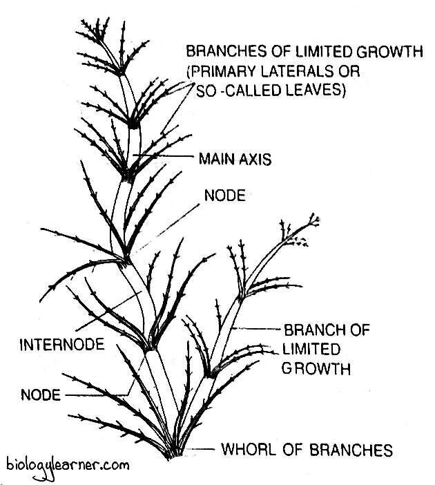
The main axis of Chara is erect, long, branched, and differentiated into nodes and internodes.
Internodes
The internode is cylindrical and consists of a single, much elongated or oblong inter nodal cell or axial cell.
The axial cells in some species may be surrounded by a one-celled thick layer called the cortex, and such species are called corticate species, e.g., C. zeylanica, C. hatei, C. fragilis, etc. The species in which the cortical layer is absent are known as ecorticate species, e.g., C. wallichii, C. corallina, C. braunii, C. succinata, etc.
The cortex is composed of a sheath of vertically elongated, narrow cortical cells. Each cortical cell is much smaller in diameter than the axial cell.
The cortical cells derive from the node. After originating, 50% of the cortical cells grow upward as the ascending filaments and the remaining 50% grow downward as the descending filaments.
The ascending filaments surround the lower half, and the descending filaments cover the upper half of the axial cell.
Nodes
The node is complex in structure and consists of a pair of central small cells surrounded by 6–20 peripheral cells. Three types of appendages are developed from each node of the main axis. These appendages are:
- Branches of limited growth or branchlets
- Branches of unlimited growth or long branches
- Stipulodes
Branches of Limited Growth
The branches of limited growth, or primary laterals, are short branches arranged in whorls on the nodes of the main axis or on branches of unlimited growth. These branches are also known as branchlets, branches of the first order, or leaves.
In a whorl, there are about 6 to 16 branchlets developed. Each branchlet is also divided into 5–15 nodes and internodes.
The nodes develop some unicellular, hair-like secondary laterals. Sex organs and stipulodes (unicellular outgrowths) are formed on the lower nodes of the branchlets.
Branches of Unlimited Growth
The branches of unlimited growth develop from the older nodes of the main axis (the axils of the branchlets). These branches are also called axillary branches or long laterals.
The axillary branches are differentiated into nodes and internodes like the main axis. Each node bears primary laterals.
Stipulodes
The stipulodes are short unicellular outgrowths developed from the lower nodes of the branches of limited growth (branchlets).
In most of the species of Chara, the number of stipulodes at each node is twice the number of branchlets. Such species are called bistipulate, e.g., C. tomentosa, C. baltica, C. burmanica, C. contraria, etc. Some species are unistipulate, e.g., C. braunii, C. coralline, and C. nuda, as the number of stipulodes at each node is equal to the number of branchlets at that node.
When the stipulodes are present in one whorl at each node, the species are called haplostephanous (e.g., C. braunii), and those with two whorls at each node are called diplostephanous (e.g., C. delicatula).
Cell Structure of Chara
The main axis of Chara is composed of mainly two types of cells: nodal cells and internodal cells.
Nodal Cells
The nodal cells are small, uninucleate, with dense granular cytoplasm. Each cell contains many discoid chloroplasts. The chloroplasts are without pyrenoids.
Small vacuoles may be present in the cytoplasm. The cell is surrounded by the cell wall, which is made up of cellulose.
Internodal Cells
The internodal cells are multinucleate (the single nucleus in the internodal cell is divided by amitosis division to form several nuclei) and much elongated. Each cell possesses a large central vacuole in the cytoplasm.
The cytoplasm is differentiated into the outer stationary ectoplasm and the inner rotatory endoplasm. Many discoid chloroplasts lie embedded in the outer exoplasm. Pyrenoids are absent in chloroplasts. The endoplasm develops a thin sieve around the large vacuole.
The endoplasm shows streaming movements. In a constant state of rotation, the endoplasm flows on one side of the vacuole and flows down on the other. The streaming of the cytoplasm is due to the alternating contraction and expansion of the protein fibrils, which remain fixed to the cell wall.
The cell wall of the internodal cell is also composed of cellulose. However, it is richly encrusted with calcium carbonate and silica, producing hardness and brittleness.
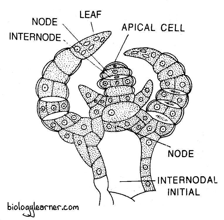
Growth of the Thallus
Growth of the thallus in Chara takes place by a single large dome-shaped apical cell, situated at the tip of the axis. The apical cell cuts off segments at its posterior surface.
Each segment divides transversely into two cells: an upper biconcave, the nodal initial, and a lower biconvex, the internodal initial.
The internodal initial does not divide further and elongates considerably to form the long internode of the axis.
The nodal initial divides by several vertical divisions and finally forms two central cells surrounded by 6–20 peripheral cells. Each peripheral cell divides into an apical initial, which later gives rise to a primary lateral and a basal nodal cell.
The basal nodal cell further divides into upper and lower cortical cells. In Chara, thus, the internode is surrounded by a cortical layer of vertically elongated rows of cortical cells.
The primary laterals grow in the same way, but the internodes are shorter than those of the main axis.
Reproduction in Chara
Chara reproduces by vegetative and sexual methods only. Due to the lack of non-motile and motile spores, asexual reproduction is absent in Chara.
Vegetative Reproduction
Vegetative reproduction in Chara occurs by various kinds of reproductive bodies which, on detachment from the parent plant, give rise to new plants.
The common methods of vegetative reproduction in Chara are as follows:
Amylum Stars
Amylum stars are star-shaped aggregates of cells develop on the lower nodes of the main axis. The cells are densely filled with amylum starch and hence are called amylum stars.
After being detached from the parent plant, the Amylum stars directly develop into new Chara plants, e.g., C. stelligera.
Bulbils
The bulbils are small oval or spherical tube-like structures that develop either on rhizoids (e.g., C. aspera) or on lower nodes of the main axis (e.g., C. baltica).
Under favourable conditions, on detachment from the plant, each bulbil germinates into a new thallus.
Amorphous Bulbils
In some species of Chara, such as (e.g., C. delicatula, C. fragifera, and C. baltica), many small cells aggregate at the lower node of the main axis or at rhizoids and form many lateral outgrowths called amorphous bulbils. These clumps of cells are laden with food materials.
The amorphous bulbils get separated and grow into new plants.
Secondary Protonema
The secondary protonema are tubular or filamentous structures developed from the primary protonema or from the basal cell of the rhizoid.
Like the primary protonema, the secondary protonema develops into Chara plants.
Sexual Reproduction
Sexual reproduction in Chara is of the advanced oogamous type. Sex organs are highly specialised and bear a superficial resemblance to the multicellular sex organs of Archigoniates (e.g., bryophytes, pteridophytes, gymnosperms). The male sex organ is called the antheridium or globule, while the female sex organ is known as the oogonium or nucule.
In Chara, most of the species are monoecious or homothallic (the male and male sex organs are borne on the same plant), e.g., C. zeylanica. Some are dioecious or heterothallic (male and female sex organs are borne on different Chara plants), e.g., C. wallichii.
Monoecious species are protandrous, i.e., the globule matures before the nucule. The sex organs develop on the adaxial side (i.e., side facing the axis) of the nodes of the branches of limited growth or primary laterals. The nucule always lies above the globule at the same node.
Globule
The globule is the male sex organ in Chara. It arises on the primary lateral.
Development of Globule
In Chara, the antheridium (i.e., globule) develops from a single superficial peripheral cell at the node of the primary lateral on the adaxial side. The peripheral cell functions as the antheridial initial.
The antheridial initial first divides transversely to form a basal pedicel cell and a terminal antheridial mother cell. The pedicel cell does not divide further and forms the pedicel or stalk of the antheridium.
The antheridial mother cell increases in size and divides by two successive vertical divisions at right angles to each other to form four cells, the quadrant (four-celled stage).
All four cells of the quadrant are then divided by a transverse division to form eight cells, called the octant stage.
Each cell of the octant divides by periclinal division and forms two layers of eight cells each. The cells of the outer or inner layer then undergo another periclinal division. As a result, three radial layers of eight cells each are formed.
The outermost eight cells enlarge laterally and form a curved plate of eight shield cells. The cells of the middle layer elongate toward the centre to form the manubrium.
The cells of the inner layer become the primary capitulum. Each primary capitulum cell divides to form two to six secondary capitulum cells. Sometimes the secondary capitulum cells divide further and form tertiary capitulum cells.
Each secondary capitulum cell divides repeatedly and forms 2 to 4 long multicellular antheridial filaments or spermatogenous filaments. The antheridial filament may be branched or unbranched and consists of 25 to 250 uninucleate cells.
The cells in the antheridial filaments function as the sperm mother cells. Each sperm mother cell metamorphoses into a single spirally coiled, biflagellated antherozoid or sperm.
Structure of Globule
The mature globule is a large, hollow, spherical, bright yellow or red structure. It is attached to the node by a large cell called the pedicle.
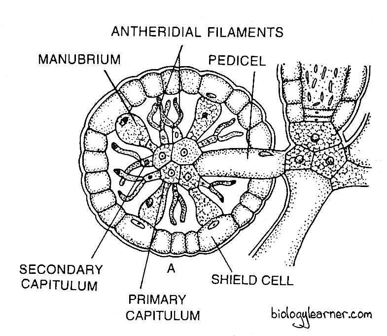
The globule has eight curved plates, the shield cells, situated towards the outer side. The eight shield cells are joined end to end, forming a pseudocellular wall structure or wall of the globule. These cells are filled with red or yellow pigments, giving the characteristic colour to the globule.
From the centre of each shield cell, a rod-shaped radially elongated structure is developed, called the manubrium or handle cell. The manubria are eight in number and project towards the centre of the antheridial cavity.
Each manubrium bears a globose terminal cell called the primary capitulum cell or primary head cell. The primary capitulum develops two to six or more secondary capitula.

In the globule, 48 or more secondary capitula are formed. Each secondary capitulum cell produces 2 to 4 long, whip-like, branched or unbranched, multicellular antheridial filaments (i.e., spermatogenous filaments).
Each antheridial filament contains 25-250 uninucleate cells. These cells are small, thin-walled, and function as spermatozoid mother cells (sperm mother cells). The spermatozoid mother cell ultimately transformed into a single, spirally coiled, biflagellate antherozoid.

Antherozoids
The antherozoid is a unicellular, uninucleate, biflagellated, spirally coiled, elongated structure. Its anterior region is called the head or orstellum. The orstellum consists of a layer of microtubules, flagella, and basal bodies.
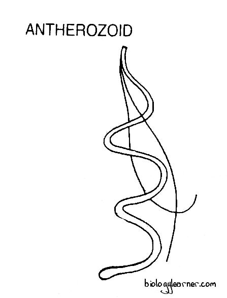
The middle region of the antherozoid is the body, and it is coiled spirally.
Liberation of Antherozoids
At maturity, the shield cells of the globule separate from each other and expose antheridial filaments in water. The antherozoids are liberated by the gelatinization of the sperm mother cells.
Nucule
The nucule is the female sex organ in Chara. Like globule, it also arises on the primary lateral.

Development of Nucule
The nucule or oogonium develops from a single peripheral nodal cell of the primary lateral. The peripheral nodal cell functions as the oogonial initial.
The oogonial initial first divides by two transverse divisions to form a row of three cells (a three-celled filament). The lowermost is the pedicel cell, the middle one is the nodal cell, and the uppermost one functions as the oogonial mother cell.
The pedicel cell remains undivided and elongates to form the pedicel or stalk of the oogonium. The middle nodal cell divides by several vertical divisions to form five sheath initials or peripheral cells surrounding a single central cell. The central cell does not divide further and functions as the node of the oogonium.
The oogonial mother cell enlarges in size and divides transversely into a lower small stalk cell and an upper large oogonium. The oogonium enlongates and its contents metamorphose into a single uninucleate egg or ovum.
The sheath initials elongate, grow upward, and divide by transverse division to form two tiers of five cells each. The five cells of the upper tier function as corona cells and develop the corona of the oogonium. The five lower tier cells form the tube cells. The tube cells elongate and spirally twist in a clockwise direction outside the oogonium.
Subsequently, the egg becomes filled with a large amount of starch and oil. The nucleus of the egg migrates towards the lower side. As a result, a receptive spot develops at the apical region of the oogonium.
Structure of Nucule
The mature nucule is a large, green, oval or elliptical structure. It attaches to the node of a primary lateral (just above the globule in monoecious species) with the help of a pedicel cell.

The nucule is remains surrounded by five long, spirally twisted tube cells. These cells function as a protective sheath around the oogonium.
Each tube cell terminates in a small erect cell at the apex of the nucule, called the corona cell. The five corona cells together form the crown or corona of the nucule.
The nucule contains a single egg or ovum densely filled with a large amount of starch and oil. In the apical region of the nucule, a receptive post is present.
Fertilization
During fertilisation, in the nucule, the spirally twisted tube cells just below the corona get separated slightly from one another and form five narrow slits or openings.

The antherozoids are attracted toward the mature egg due to the chemotactic response. Through the openings, the antherozoids enter into the sheath of the nucule.
Many antherozoids penetrate the gelatinized wall of the nucule. Only one antherozoid enters the egg at the receptive spot and fertilises the egg to form a diploid zygote.
Zygote or Oospore
After fertilization, the zygote secretes a thick wall around itself to form an oospore. Oil globules accumulate in the protoplast.
The oospore is a hard, spherical or elliptical structure. It may be black (e.g., C. corallina), brown (e.g., C. inferma), or light yellow (e.g., C. flauda).
The oospore is retained for some time within the oogonium and the oogonial wall as well as the oospore walls become thick. The oospore nucleus(2n) moves towards the apical region.
Ultimately, the oospore is liberated from the oogonium by the decay of the sheath (oogonial wall). It rests on the mud at the bottom of the pond and remains dormant for a few weeks or more.
Germination of Oospore
After a period of rest, the oospore germinates into a new Chara plant under favourable conditions.
The diploid oospore nucleus divides by meiosis to form four haploid daughter nuclei. The oospore is now divided by a transverse wall (septum) into two unequal haploid cells.
The upper smaller apical lenticular cell contains a single nucleus, and the large basal cell contains the remaining three nuclei. The three nuclei of the basal cell degenerate gradually.
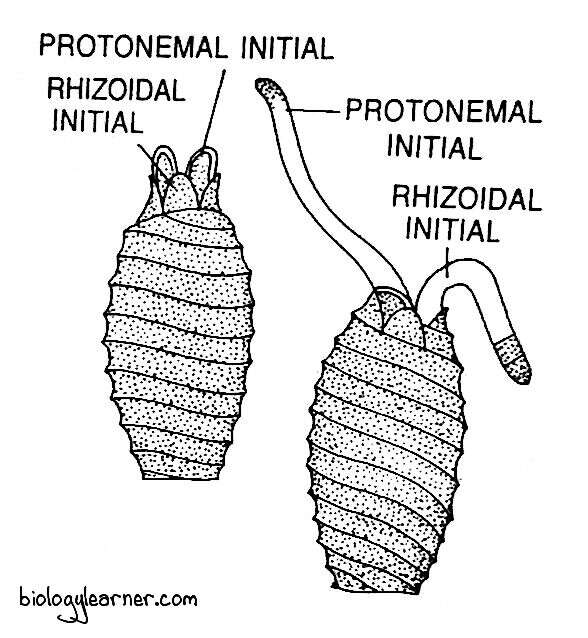
The lenticular cell is exposed by the ruptures of the oospore wall. Subsequently, it divides mitotically by an oblique longitudinal wall to form a rhizoidal initial and a protonemal initial. Both the initials grow into knob-like structures in the opposite direction.

The rhizoidal initial elongates and develops into a colourless filamentous rhizoid. The protonematal initial forms an erect, green epiterranean primary protonema.
The primary protonema is differentiated into nodes and internodes. The mature Chara plant arises as a lateral branch from the protonema.

