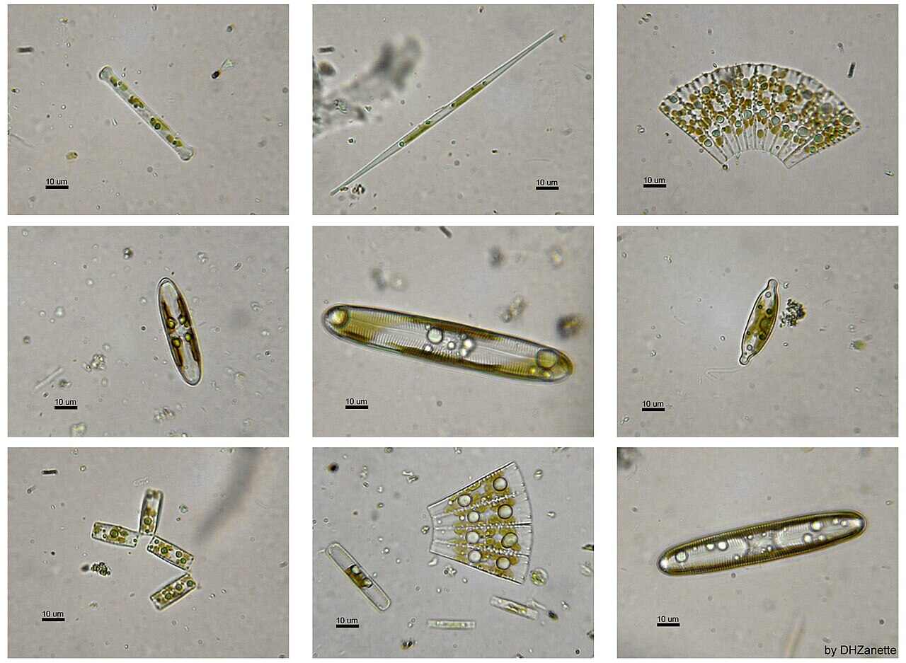
Algae present in the group Bacillariophyta (class Bacillariophyceae) are known as diatoms. They constitute a very large assemblage of unicellular algae.
Diatoms differ from other algae due to their symmetrical structure and delicately designed cell walls. Thus, diatoms are considered the most beautiful microscopic algae.
Characteristics of Bacillariophyta
- Diatoms are found in freshwater as well as marine habitats. Some members are attached to the substrata, while others are free-floating.
- The plant body is unicellular or sometimes colonial. It is always diploid.
- The thalli are of different shapes. They may be oval, spherical, triangular, rod-shaped, disc-shaped, or boat-shaped.
- The cells are bilaterally or radially symmetrical.
- All vegetative cells are diploid.
- The cell wall of diatoms is called the frustule. It consists of two overlapping valves, or halves, arranged in the form of a box with its lid.
- The wall is made up of pectic substances with a high amount of silica deposit. It shows characteristic secondary structures like spines and bristles.
- Cells usually possess several discoid or two large plate-like chromatophores, which contain many photosynthetic pigments.
- Photosynthetic pigments are chlorophyll-a, chlorophyll-c, and carotenoids, along with xanthophylls like diatoxanthin, diadinoxanthin, and fucoxanthin. Diatomin (xanthophylls + carotenoids) gives the thallus a golden brown colour.
- Pyrenoids are present.
- Reserve food materials are in the form of fats, volutin, and chrysolaminarin.
- Some cells exhibit gliding movement caused by the streaming of cytoplasm.
- The only motile structure formed in this group is the male gamete of the centric diatoms. It has a single pantonematic flagellum, which is apically inserted.
- Reproduction usually occurs through cell division and auxospore formation.
- Sexual reproduction takes place by either anisogamy or oogamy.

Distribution of Bacillariophyta
There are about 250 genera of living diatoms, with around 100,000 species.
Diatoms are found in all possible habitats, especially in freshwater and marine water. They are usually associated together in great abundance in the aquatic ecosystem.
Some diatoms are either strictly freshwater (e.g., Navicula pupula, Melosira variens, Denticula tenuis, Synedra, Asterionella, etc.) or strictly marine (e.g., Fragilaria, Corethron, Sceletonema, Biddulphia, etc.). A few members, such as Frustulia, Pinnularia, and Navicula, are terrestrial.
Cymbella and Gomphonema occur as epiphytes on other algae like Cladophora and Enteromorpha. Licmophora is an endozoic member, growing within living organisms.

Plant body of Bacillariophyta
Diatoms are unicellular. The cells are of different shapes. They may be circular, oval, elongated, triangular, boat-shaped, disc-shaped, wedge-shaped, straight rod-like, etc.
Sometimes diatoms form groups and are embedded in a gelatinous matrix. In colonial form, the cells may be present as a branched body (Licmophora) or in chains (Melosira, Cymbella).

Cell Structure of Bacillariophyta
Structurally, the diatom cell consists of two parts: the cell wall and the protoplast.
Cell Wall
The cell is covered by a siliceous wall called the frustule. It is differentiated into an inner layer and an outer layer.
The inner layer is a thin, continuous pectin membrane, which encloses the protoplast. The outer layer consists of two overlapping halves, one of which fits the other like the lid of a pill box or like the two parts of a petri dish.
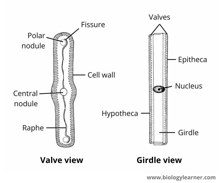
The upper half is called epitheca, and the inner half is called hypotheca. Each theca is made up of a valve (the main surface) and an incurved margin, known as the connecting band or cingulum.
The two connecting bands in the diatom cell are firmly united in the overlapping region, which is termed the girdle. Thus, a diatom cell can be seen in the following two different aspects when viewed from the surface:
- Valve View: It is called the top view. In this view, the valve side is uppermost.
- Girdle View: In this case, the overlapping band or girdle is uppermost. It is also known as the side view.

The mucilage material covering the cell wall is composed of pectin substances impregnated with silica. Due to the presence of silica, various kinds of ornamentation, such as ridges, pits, and fine pores, are confined to the valve portion of the cell wall. The fine working on the ridges on the valves is called striae.
The striation on the valves is arranged in two patterns: pennate type and centric type.
Pennate Type
The striations are arranged in a pennate manner and have raphae (elongated slits), pseudoraphae, central nodules (thickenings of the wall), and polar nodules (thickenings present at the ends).
These types of diatoms are placed in the order Pennales and are commonly known as pennate diatoms. They are bilaterally symmetrical. E.g., Pinnularia, Navicula.

Centric Type
The striations are arranged radially. There are no raphae. The diatoms are placed in the order Centrales and are commonly called centric diatoms. These are radially symmetrical. E.g., Melosira, Cyclotela.
Protoplast
The protoplast is differentiated into a plasma membrane, cytoplasm, and a single nucleus with several chromatophores.
The plasma membrane encloses a large central vacuole surrounded by cytoplasm. The cytoplasm contains mitochondria, golgi bodies, and endoplasmic reticulum.
In centric diatoms, many discoid chromatophores are seen. But there are one or two chromatophores in pennate diatoms.
The chromatophores are olive-green to yellowish-brown. They contain chlorophyll-a and chlorophyll-c, which are masked by other pigments such as β-carotene and xanthophylls (fucoxanthin, diatoxanthin, diadinoxanthin, etc.). Pyrenoids are usually absent.
Chrysolaminarin and oil droplets are present as reserve food materials.
Movement
The centric forms are non-motile. Many pennate diatoms, in particular those with a raphe, exhibit slow, spontaneous gliding movements.
The gliding movement is caused by the streaming of cytoplasm, by circulation within the raphe, and by the release of mucilage. The rate of movement varies from 0.2 to 25 μm/s (Prescott, 1969).
The movement takes place only in the forward and backward directions along the longitudinal axis.
Reproduction in Bacillariophyta
Members of Bacillariophyta reproduce by vegetative and sexual methods. Asexual reproduction by the formation of non-motile or biflagellate zoospores for some planktonic oceanic species of the Centrales has been claimed by some workers, but there is still doubt as to whether such spores are actually formed in diatoms.
Vegetative Reproduction
In diatoms, vegetative reproduction takes place through cell division. Cell division usually occurs at midnight or in the early morning. Also, the presence of aluminium silicate in water stimulates the process.
During cell division, the protoplast increases in diameter. The cell enlarges in volume, overlapping theca become loose, and the girdle gets separated.
The nucleus divides mitotically in a plane perpendicular to the valves and forms two daughter nuclei. Now the protoplast undergoes longitudinal division in a plane parallel to the valves, resulting in the formation of two daughter protoplasts.
One half of the protoplast (i.e., the daughter protoplast) remains in epitheca and the other one in hypotheca. Thus, one side of the protoplast remains naked.
Each half of the protoplast now secretes a new siliceous valve on its naked side. The newly formed valves always behave as hypotheca, while the older theca (epitheca and hypotheca of the parent cell) become the epitheca of the daughter cells.
Finally, connecting bands are developed between the theca, and the daughter cells get separated from each other.

Of the two daughter cells that are formed, one has the same size as the parent cell, and the other is reduced in size (the side where the hypotheca behaves as the epitheca in the parent cell).
Thus, in a population of diatom cells, during cell divisions, there is normally a progressive decrease in the average cell size (as some cells gradually become reduced in size). It is known as the MacDonald-Pfitzer law.
