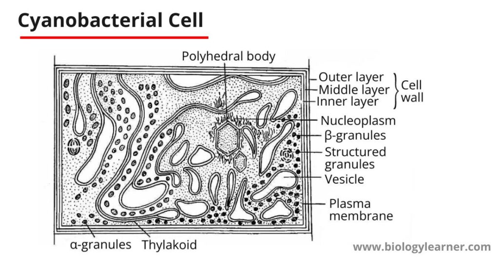
Cyanobacteria are the most primitive types of organisms (plants) with simple prokaryotic cellular structures.
With the help of an electron microscope, Pankratz and Bowen (1963) studied the structure of cyanobacterial cells. The detailed cell structure of cyanobacteria is as follows:
Cell Sheath
The cyanobacterial cell is surrounded by a hygroscopic gelatinous sheath, which is made up of three layers of microfibrils. The microfibrils are composed of pectic acids and mucopolysaccharides. Sheaths are reticulately arranged within a matrix to give a homogeneous appearance.
The function of the cell sheath is to absorb and retain water.

Cell Wall
A multi-layered wall is present below the gelatinous sheath. The cell wall is made up of four layers which are designated numerically as LI, LII, LIII, and LIV.
The layers LI and LIII are electron transparent, while LII and LIV are electron-dense.
- Layer LI: The innermost layer of the cell wall is LI. It is present next to the plasma membrane. Layer LI is of about 3-10 nm thickness and is enclosed by layer LII.
- Layer LII: LII is an electron-dense, thin layer. It is about 10-1000 nm in thickness. The layer LII consists of muramic acid, glutamic acid, alanine, glucosamine, and α-di-amino-pamelic acid.
- Layer LIII: The electron transparent Layer III is of about 3-10 nm thickness, just like layer LI.
- Layer LIV: The outermost layer is LIV which is a thin and again electron-dense layer.
Plasma Membrane
Below the cell wall, a thin cytoplasmic bilayer membrane called the plasma membrane is present. The plasma membrane consists of two electron-opaque layers separated by a translucent layer. It has a thickness of 7 nm, is selectively permeable, and maintains the physiological integrity of the cell.
Cytoplasm
The cytoplasm is differentiated into two regions, the outer colored peripheral region which is called the chromoplasm, and the central colourless region called the centroplasm or central body.
Chromoplasm
The chromoplasm contains flattened vesicular structures called photosynthetic lamellae or thylakoids.
Lamellae
In the peripheral part of the cyanobacterial cell (in chromoplasm), many lamellae or thylakoids are present. The lamellae are arranged in two or more parallel stacks or scattered irregularly. Each lamella is represented by a flat sac, which is surrounded by a unit membrane. Each unit membrane is about 75 Å in thickness.
These lamellae are separated by a space of 50 μ. This space is occupied by many rows of discoidal phycobilisomes, which contain phycocyanin pigment and, to a lesser extent phycoerythrin pigment.
Centroplasm or Central Body
The central body, or centroplasm, is colorless and irregular in shape, occupying more or less a quarter or one-third of the total volume of the cell.
Some people are of the opinion that the centroplasm is the storehouse of food and according to others, it is an incipient nucleus or nucleoplasm.
The nucleoplasm contains numerous fine, randomly oriented DNA fibrils.
Cytoplasmic Inclusions
In a typical cyanobacterial cell, many sub-cellular organelles such as cyanophycean granules, gas vacuoles, polyglucoside bodies, ribosomes, polyhedral bodies, polyphosphate bodies, etc. are present.
- In many cyanobacteria (e.g., Anabaena, Gloeotrichia, Microcystis, Oscillatoria, etc.), pseudovacuoles (pockets of gas) are found to be distributed throughout the cytoplasm. These pseudovacules provide greater buoyancy in such species and also act as light screens against intense illumination.
- The proteins present in the cell are synthesized by ribosomes.
- Cyanophycean granules or cyanophycin store reserve food material.
- Polyphosphate bodies are meant for the storage of phosphorus.
- Polyhedral bodies are also called carboxysomes. They produce an important enzyme called ribulose diphosphate carboxylase.
- Other granules are called storehouses of nitrogen.

This topic is well-organized…
Way cool! Some very valid points! I appreciate you penning this post plus the rest of the site is also really good.