The female gametophyte of gymnosperms is a multicellular structure serving the functions of bearing the archegonia and nourishing the developing embryo. It exhibits a progressive series of significant changes leading towards angiosperm conditions.
Due to the absence of fossil records, our knowledge is obtained from extant forms, which do not represent a series but the ends of many series.
Development of Female Gametophyte in Gymnosperms
Before discussing evolution, a brief account of the different stages of development of the female gametophyte is outlined below:

Cycas
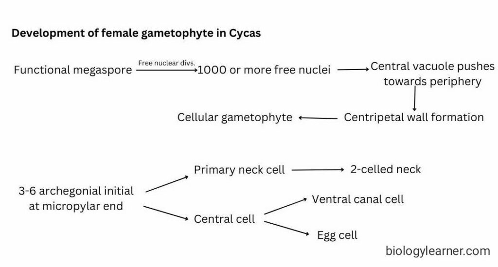
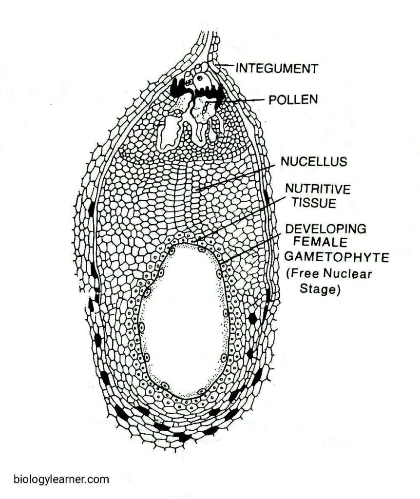
In Cycas, the female gametophyte is characterized by the free nuclear stage, the wall formation stage, and the archegonium development stage.

- The gametopgyte is monocarpic (i.e., out of the four megaspores, only one becomes functional) and ebdosporic, and the number of free nuclei following mitosis is more than 1000.
- Wall formation is centripetal, with the endosperm of larger cells at the chalazal end and smaller ones towards the micropylar end.
- The number of archegonia is between 2-8, the neck canal cell is completely absent, and the venter is surrounded by a nutritive jacket layer.

Pinus
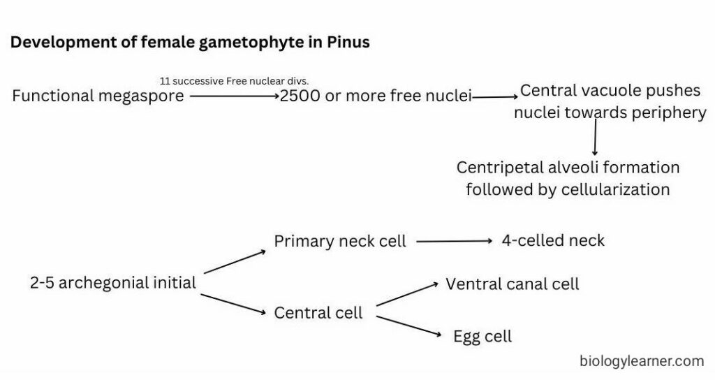
In Pinus, the gametophyte is also characterized by the three stages of development, i.e., the free nuclear stage, the wall formation stage, and the archegonium development stage.
- The gametophyte is monosporic and endosporic, and the megaspore starts development after pollination. There is an interval of 13 months between the origin of the megaspore and the development of the mature prothallus.
- Free nuclei are not more than 2500. Wall formation begins at the micropylar end and extends centripetally at the base.
- Alveoli are formed with many nuclei in each chamber, and the gametophyte becomes cellular.
- The number of archegonia is 2-4, and the cytoplasm of the egg is fibrillar. A distinct jacket layer surrounds the central cell. Neck canal cells are not formed.

Ginkgo
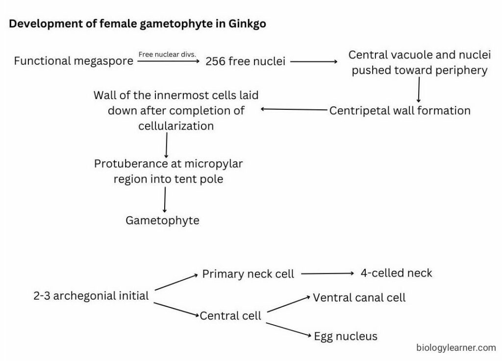
Here, the female gametophyte is also characterized by the three stages of development: the free nuclear stage, the wall formation stage, and the development of archegonium.

- The gametophyte is monosporic and endosporic. The megaspore enlarges after pollination.
- The number of free nuclei is 256, and a peripheral thin membrane is secreted internal to the megaspore wall.
- Outer cells are completely surrounded by cell walls, but the innermost cells have no wall on the inside. Inner cells secrete their own walls.
- Cells in the central region of the gametophyte are not joined by the common wall.
- A beak is present at the micropylar end, called the tent pole, around which a circular crevice develops. Due to the presence of chloroplasts, all cells are green.
- Two or rarely three archegonia are present.

Gnetum
In Gnetum, the gametophyte is characterized by only two stages: the free nuclear division and the wall formation stage.
- Here, the gametophyte is tetrasporic and endosporic. The number of free nuclei varies from 256 to 1500, depending on species.
- Spathulate at the chalazal end, and a large central vacuole is present.
- Wall formation begins at the chalazal end and becomes cellular. But the micropylar end remains free nuclear.
- Gametophyte is partly cellular and partly free nuclear.
- Archegonia are absent. A few free nuclei at the chalazal end function as eggs.

Discussion
A general sequence in the early development of the female gametophyte shows a free nuclear stage followed by cellularization by anticlinal walls. The number of free nuclear divisions is fixed and depends on the size of the gametophyte.
The following evolutionary tendencies are noticed in the genera studied that ultimately lead to a stage in Gnetum:
- Inequality in the time of archegonium or the formation of egg
- Elimination of the venter canal cell
- Distribution of archegonia
- Delayed endosperm formation
Inequality in the time of Archegonium or the Formation of Egg
In the most primitive condition of the gametophyte, the egg does not appear until the endosperm is nearly fully grown, as in Cycas and Ginkgo.
In Coniferales (e.g., Pinus), all stages are found except the one just preceding the elimination of archegonium, which is attained in Gnetum.

The extreme case is exhibited in the genus Gnetum, where the egg matures in the most embryonic stage of the gametophyte (at the free nuclear division stage).
Elimination of the Venter Canal Cell
The general tendency of the archegonia among the gymnosperms is to eliminate the venter canal cell.
The walled venter canal cell is retained in Ginkgo and Pinus.
In other groups, the wall has disappeared, and the venter canal cell is represented by a free nucleus. Its presence in ginkgo is a safe inference to conclude that the venter canal cell was present among the ancient gymnosperms.
Distribution of Archegonia
It is evident that the position of the archegonia is related to the position of the pollen tube, which sometimes reaches the embryo sac before the archegonial initials are selected.
The tendency of archegonia to become definite in number is seen and the archegonia are then organized in two ways.
- Either as individual archegoium, each with its own jacket and chamber.
- As an archegonial complex with a common jacket and chamber (e.g., Cupressaceae).
From these two were derived the free eggs of Gnetum, where archegonia are eliminated from the ontogeny.
Delayed Endosperm Formation
In all genera except Gnetum, the female gametophyte functions as storage tissue or endosperm, whereas in Gnetum, endosperm is delayed until fertilization.

Conclusion
The above discussions clearly demarcate the advanced tetrasporic condition from the other monosporic gametophytes.
The elimination of the neck canal cell, then the wall around the venter canal cell, and finally the archegonium are the few important events in female gametophyte evolution.
