The cardiac cycle refers to the sequence of events that results in the continuous and rhythmic contraction and relaxation of the heart chambers.
All the events in a cardiac cycle occur in one heartbeat. It involves a complete contraction (systole) and relaxation (diastole) of the atria and ventricles, ensuring efficient blood circulation through the arteries and veins in a synchronized manner.
Each cardiac cycle, or heartbeat, takes approximately 0.8 seconds to complete. It is called cardiac cycle time.

Phases of Cardiac Cycle
The human cardiac cycle consists of four main phases. These are-
- Atrial Systole
- Ventricular Systole
- Atrial Diastole, and
- Ventricular Diastole
In addition to these four main phases, there is an intermediate phase known as protodiastole that marks the end of systole and the beginning of diastole.

Atrial Systole
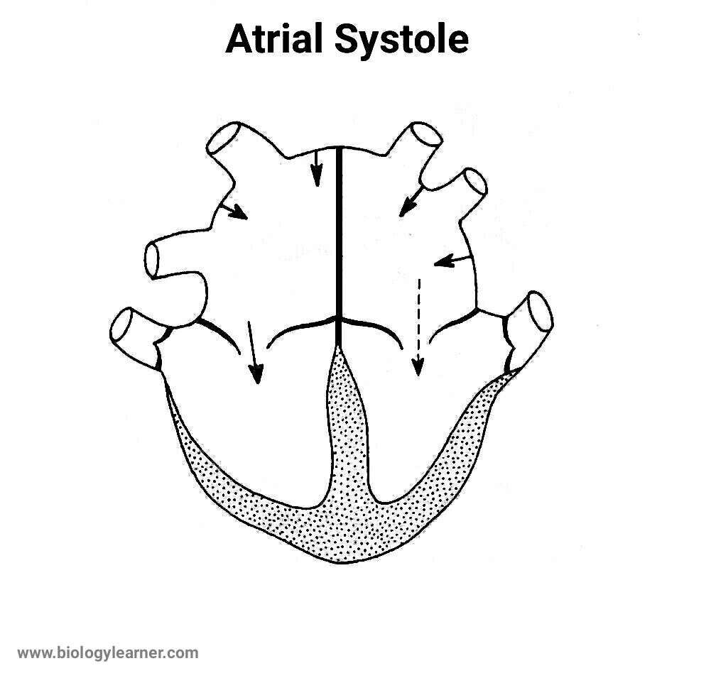
The atrial systole initiates the cardiac cycle. This phase is also called presystole, the last rapid phase, or atrial kick.
During atrial systole, the atria contract and pump the blood out of the atria to the ventricles. The left atrium (LA) contracts a little after the right atrium (RA), as it is slightly away from the natural pacemaker or sinoatrial (SA) node.

The cardiac impulse originating in the SA node is transmitted through the internodal tract to the atrial walls, which triggers the contraction of the atrial walls. This phase is completed in about 0.10 seconds.
Ventricular Systole
Ventricular systole begins immediately after the end of atrial systole.
The purkinje fibers relay the impulses to the ventricular walls, resulting in the contraction of the ventricles. This phase usually lasts for about 0.3 seconds.

The ventricular systole can be further divided into the following sub-phases:
1. Isovolumetric Ventricular Contraction
The isovolumetric ventricular contraction is the first stage of the ventricular systole. At this stage, the muscle tension increases rapidly, and the ventricles begin to contract without any change in volume.
During isovolumetric contraction, the pressure inside the ventricle increases. When this pressure exceeds the atrial pressure, both the atrioventricular (AV) valves (the mitral valve and the tricupsid valve) are closed. The two semilunar (SL) valves, the aortic valve and the pulmonary valve, also become closed. So, the blood still remains in the ventricle.
As a result, the volume of the ventricles remains the same while their internal pressure increases rapidly. This phase takes about 0.05 seconds to complete.
2. Maximum Ventricular Ejection
The SL valves open due to pressure differences in the ventricles during this stage. At high pressure, approximately 70% of the ventricular blood is pumped out from the ventricles through the aorta and the pulmonary arteries.
The pressure inside the left ventricle is higher (about 80 mm of Hg) than that in the right ventricle (about 8 mm of Hg). Hence, the blood is rushed more forcefully into the systemic aorta compared to the main pulmonary artery. This phase lasts for about 0.11 seconds.
3. Reduced Ventricular Ejection
In this stage, some of the ventricular muscle fibers begin to relax, and the remaining 30% of the blood is slowly ejected from the ventricles into the aorta.
The intraventricular pressure also starts decreasing slowly. This phase continued for about 0.14 seconds.
Protodiastole
It is the intermediary stage between the end of the systole and the beginning of the diastole.
After reducing the ventricular ejection, the ventricular pressure decreases and becomes lower than the blood pressure inside the major arteries.
The duration of this phase is about 0.04 seconds.
Atrial Diastole
The atrial diastole is the stage when the atria are relaxed and filled with blood, and it begins immediately before the ventricular diastole phase.
The AV valves are closed during this stage. The two vena cava, i.e., the superior and inferior vena cava, bring the deoxygenated blood to the right atrium. Simultaneously, the pulmonary veins carry the purified or reoxegenated blood to the left atrium.
This phase occurs while the ventricles are contracting (ventricular systole) and takes 0.7 seconds to complete.
Ventricular Diastole
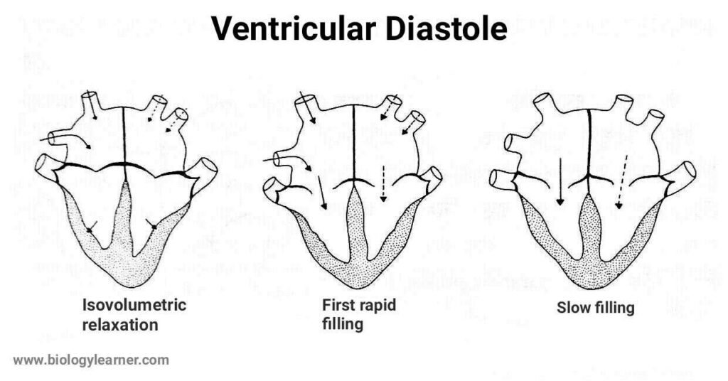
It is the stage when the blood is transmitted to the ventricles from the atria, increasing the ventricular pressure and volume. The duration of this phase is about 0.5 seconds.

The ventricular diastole is divided into the following four sub-phases:
1. Isovolumetric Ventricular Relaxation
Volumetric ventricular relaxation is the initial phase of ventricular diastole. During this stage, the muscle tension of the ventricular wall decreases without any change in the volume of the ventricles.
The SL valves become completely closed due to the reduced pressure. The AV valves that close at the beginning of ventricular systole are still closed at this phase. So the blood cannot enter the ventricles, and the volume of the ventricles remains constant. This phase is completed in about 0.08 seconds.
However, the rapid pressure drop causes a subsequent rise in pressure within the atrium. This will lead to the next stage.
2. Rapid Ventricular Filling
At this stage, the AV valves open, and about 70% of the blood rapidly enters from the atria into the ventricles.
This phase usually lasts for about 0.09 seconds.
3. Slow Ventricular Filling
The flow of blood from the atria into the ventricles is slowed down during this stage. Approximately 20% of the atrial blood enters the ventricles.
This phase is also known as diastasis and takes about 0.19 seconds to complete.
4. Last Rapid Ventricular Filling
It is the final phase of ventricular diastole and corresponds to the atrial systole phase.
At this stage, the flow of atrial blood into the ventricles becomes rapid again (due to atrial contraction), and the remaining 10% of the blood is passed to the ventricles.
This phase usually lasts for about 0.10 seconds.

Summary of Cardiac Cycle
The relationship between the duration of various events in a cardiac cycle at a normal pulse rate is given in the following table.
I. Atrial events (0.8 seconds):
a. Atrial systole (0.1 seconds)
b. Atrial diastole (0.7 seconds)
II. Ventricular events (0.8 seconds):
a. Ventricular systole (0.3 seconds)
| Phases of Ventricular systole | Duration |
|---|---|
| i. Isovolumetric contraction | 0.05 seconds |
| ii. Maximum ejection | 0.11 seconds |
| iii. Reduced ejection | 0.14 seconds |
b. Ventricular diastole (0.5 seconds)
| Phases of Ventricular diastole | Duration |
|---|---|
| i. Protodiastole | 0.04 seconds |
| ii. Isovolumetric relaxation | 0.08 seconds |
| iii. Rapid ventricular filling | 0.09 seconds |
| iv. Slow ventricular filling | 0.19 seconds |
| v. Last rapid ventricular filling | 0.10 seconds |
