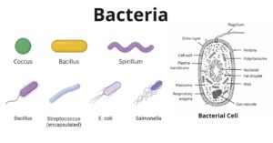
Bacteria are unicellular. Their structure is a very simple type. Bacteria are prokaryotes because they do not have a well-formed nucleus. A typical bacterial cell is structurally very similar to a plant cell.
The cell structure of a bacterial cell consists of a complex membrane and membrane-bound protoplast. There are also cell walls, cytoplasm, and nucleoids (genetic material) are present in the bacterial cell.
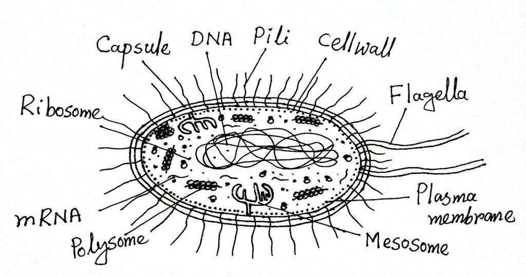
Parts of a Typical Bacterial Cell
The different parts of a typical bacterial cell are described below:

Bacterial wall
The outermost part of a bacterial cell is a bacterial wall. The bacterial wall consists of mainly two layers: the slime layer and the cell wall.
Slime Layer and Capsule
There is usually a thick slippery slime layer present just outside the cell wall of a bacterial cell. When the layer is hard, called the capsule. It is composed mainly of polysaccharides, glycoproteins, and polypeptides.
Functions of Slime Layer or Capsule
- Capsules are effective in nitrogen fixation.
- The capsule is capable of absorbing water vapor from the air.
- The capsule protects the bacteria from dryness and antibodies.
- It protects bacteria from phagocytosis and viral infections.
- The slime layer protects bacteria from toxic chemicals.
Cell wall
The thick erect elastic membrane that lies beneath the slime layer outside the bacterial cell is called the cell wall. Its thickness is around 10-25 nm and is made up of proteins, lipids, and carbohydrates. Usually, the cell wall does not contain cellulose.
The cell wall is composed of basically mucopeptide or peptidoglycan.
- In the glycan part of the peptidoglycan, N-acetyl glucosamine and N-acetyl muramic acid chain are present.
- There are two types of peptides are present in the peptide part of peptidoglycan. Like – stem peptide and linker peptide. Stem peptides is containing L-alanine, D-glutamic acid, Meso-diaminopimelic acid, and D-alanine. It is connected with the N-acetyl muramic acid of the glycan part. on the other hand, the linker peptide consists of amino acids like glycine, serine, alanine, or threonine and connects between two stem peptides.
- In Gram-positive bacteria, teichoic acid and simple polysaccharides are also present in addition to glucose in the cell wall.
- While lipoprotein, lipopolysaccharide, and phospholipids are present with peptidoglycans in the cell wall of gram-negative bacteria.
Though cell wall and chemical structure are different in gram-positive and gram-negative bacteria.
In gram-positive bacteria, the cell wall is thick and homogeneous but the cell wall of gram-negative bacteria is relatively lesser thick and heterogeneous.
Functions of Bacterial Cell Wall
- The cell wall is responsible for the shape of the bacterial cells.
- The cell wall protects the protoplasm.
- It is permeable and allows solutions to reach the protoplasmic surface.
- Plays a special role in the mechanism of bacterial transformation.
- Proteins of the cell walls are enzymatic and responsible for the synthesis of capsular polysaccharides and extracellular metabolic products.
Cytoplasmic Membrane
This is the boundary layer of the protoplast and is about 6-10 nm thick. The cytoplasmic membrane separates the cell wall from the protoplast. The main chemical components are lipids and proteins with small amounts of carbohydrates.
Electron microscopic studies appear a triple-layered structure consisting of two electron-dense layers surrounding an electron transparent layer.
Functions of Cytoplasmic Membrane
- The plasma membrane protects the protoplasm from an unfavorable environment.
- It allows the entry and exit of some specific molecules. Because of the permeable nature of the plasma membrane.
- Bacterial cell membranes contain enzymes for respiration and other metabolic processes.
- The presence of the enzyme permease plays an important role in the transport of inorganic ions, carbohydrates, amino acids, and vitamins.
Protoplast
All the lively internal part of the bacterial cytoplasmic membrane is called the protoplast. Bacterial protoplasts consist mainly of cytoplasm and nuclear bodies.
Cytoplasm
The cytoplasm is the homogeneous colloidal part of the protoplast. Bacterial cytoplasm contains imaginations of the cytoplasmic membrane in the form of mesosomes, cytoplasm also contains ribosomes. Other membrane-bound cell organelles such as mitochondria, Golgi bodies, endoplasmic reticulum, etc. are absent in the bacterial cytoplasm.
Intracytoplasmic inclusions occur in the form of volutin granules, crystals, lipids, polysaccharides, vacuoles, etc. Volutin granules consist of polymetaphosphates. Contains polysaccharides as storage products. Vacuoles are fluid-containing cavities separated from the cytoplasm by a membrane.
Ribosome
Ribosomes are small membraneless, ribonucleoprotein particles of about 10-30 nm in diameter. Most of the part of the cytoplasm is occupied by the ribosomes. There are two types of ribosomes in bacterial cells – fixed type and free type. Fixed-type ribosomes are attached to all membranes and free-type ribosomes lie free in the cytoplasm.
The sedimentation value of bacteria is in the 70s. So, the ribosome of bacteria is 70s type. There are 10000 to 15000 ribosomes in a bacterium cell. The main function of ribosomes is to take part in protein synthesis.
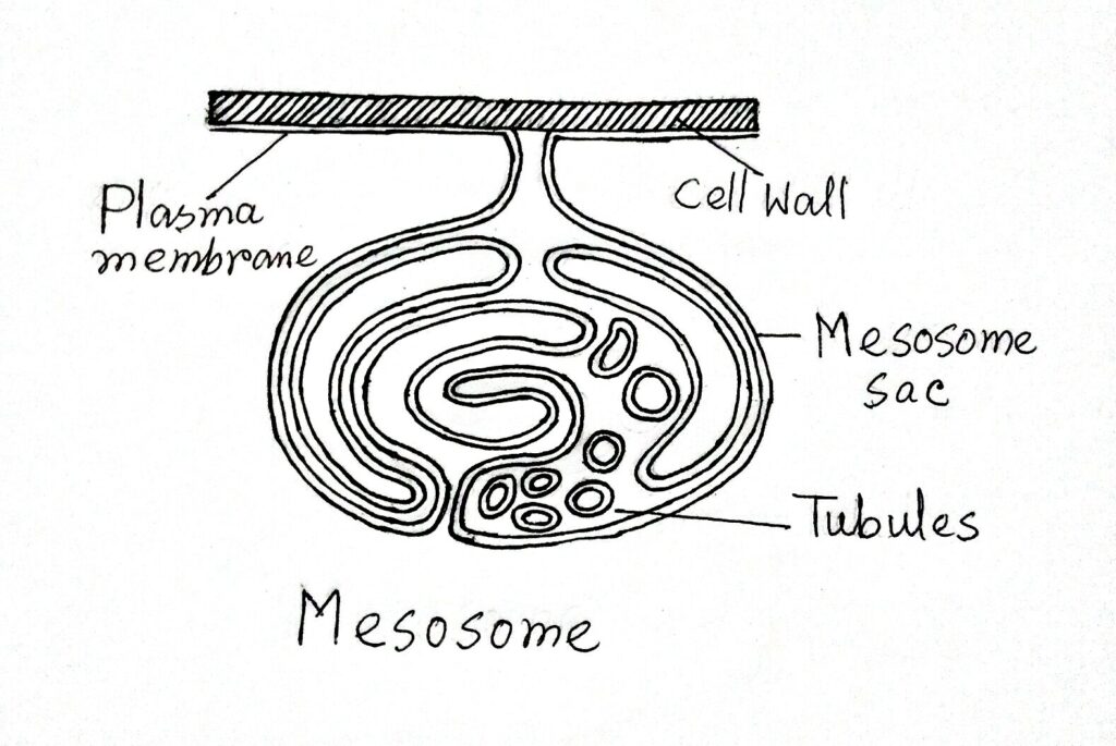
Mesosome
Mesosomes have been defined as vesicular, lamellar, or tubular pockets of membrane-enclosed by the imaginations of the cytoplasmic membrane. Previously they were called the peripheral body or chondroid. In gram-positive bacteria, mesosomes are more prominent.
According to some authors, mesosomes are simply preparation artifacts.
Functions of Mesosome
- The function of the mesosome is still unknown.
- However, according to many scientists, mesosome is a precursor to mitochondria because it contains an enzyme called cytochrome oxidase.
- Mesosome in bacteria is involved in cell wall synthesis and the formation of septa during cell division (J.Peberdy,1980).
Lamellae and Vesicles
There are single membrane-bound tube-like lamellae and spherical vesicles inside the cytoplasm of the photosynthetic bacterial cell. They are usually 30-60 mμ in diameter. They contain enzymes and pigments.
Their main function is to help photosynthesis.
Reserve Substances
The reserve substances of bacterial cells are mainly polysaccharides, glycogen, lipid granules, volutin granules, proteins, etc. These substances are floating state in the cytoplasm.
Nuclear Body
It is referred to as nucleoid and is of primitive type. Nuclear material consists of loops of supercoiled DNA together with about 10% RNA and 10% protein (mostly RNA polymerase).

With the help of an electron microscope, this body shows a diffuse area containing fibrous material without any limiting membrane. There is a double-stranded DNA forming a single circular chromosome in each nucleoid, usually 1-2 mm in length.
According to Cairns (1963), DNA is a double-stranded single circular i.e, ring-like structure 1000 mμ long, which remains diffused throughout the cell’s cytoplasm – hence such type of genetic material is called genophore. The nucleoid has the ability to replicate and mutate.
Plasmids and Episome
Plasmids are small circular DNA molecules – they replicate in the cells independently of the chromosomes. These fragments independent of chromosomes are termed plasmids. The plasmids normally contain about 2% of total genetic information and multiply independently of the chromosome.
Those plasmids which attach themselves to the chromosome are called episomes.
Surface Appendages
These appendages are arising from the surface of the bacterial cell wall. These are mainly three types: Flagella, Fimbriae or pili, and Spinae.
Flagella
Flagella are long fine hair-like appendages arising from the surface of the bacterial cell walls.
The majority of bacteria are flagellate. Flagella are 10-15 μm in length and 20-30 μm in diameter. These are made up of flagellin protein. Protein is present in spiking form. It is a single fibril.

Bacterial flagella consist of three parts: a basal body, hook, and filament.
Types of Bacteria based on Flagella
Bacteria are of the following types based on flagella:
- Atrichous: Bacteria without flagella
- Monotrichous: These bacteria are having single flagella at one pole
- Amphitrichous: Bacteria have one flagellum at each end
- Lophotrichous: Bacteria have a cluster of flagella at one or both ends
- Peritrichous: These bacteria are surrounded by flagella.
Flagella help in the movement of bacteria.
Pili or Fimbriae
Fimbriae or pili are thin and fine hair-like non-flagellar surface appendages found in some gram-negative bacteria. They look superficially similar to flagella and are shorter and thinner than flagella – hence they are also called pili. Pili are 3-25 nm in diameter and 0.5 – 20 μm in length.
Pili are two types Common pili and Sex pili.

Common pili are involved in adhesion between themselves. Sex pili are longer than the other pili and help in sexual recombination.
Spinae
These have been reported to occur in some gram-positive bacteria. Spinae are tubular pericellular, non-prosthecate rigid appendages made up of single protein moiety spinin.
The function is to adjust cells to some environmental conditions such as salinity, temperature, etc.


It’s helpful